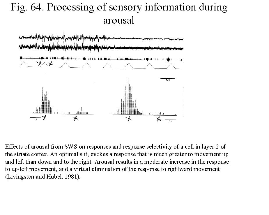
SUBCORTICAL
MODULATORY SYSTEMS.
Foundations
(Zaborszky 11-14 and 12-03, 2002)
Over the past 50 years, the basic
mechanisms of sleep-wake states have been studied with an interdisciplinary approach
embracing neurophysiology, neuroanatomy and neurochemistry. Early studies
employed lesions and stimulation of the brain to identify regions and delineate
neural systems that are involved in the generation and maintenance of
wakefulness and sleep. Such
experimental studies were also important in identifying the neuroanatomical
substrates of coma and the extreme sleep perturbations that occur in
association with brain lesions in humans. Neurophysiological research has
employed recording of single neurons in the brain to discriminate putative
wake- and sleep-generating neurons and to understand the cellular mechanisms of
sleep-wake state generation. Over the past 25 years, research has focused on
the involvement of specific neurotransmitters and corresponding chemically
specific neuronal circuits in the generation of sleep and wakefulness.
Brain Electrical Activity
During Waking and Sleep States
The states of wakefulness and sleep are
characterized by a set of three cardinal physiological correlates: brain wave
activity (electroencephalogram, or EEG), eye movements, and muscle tone.
The
background electrical activity of the brain in unanesthetized animals was
described in the 19th century, but it was first analyzed in a systematic
fashion by Hans Berger in the late twenties in the last century, who introduced
the term electroencephalogram (EEG) to denote the record of the
variations in potential recorded from the brain. The EEG can be recorded with
scalp electrodes through the unopened skull or with electrodes on or in the
brain. The term electrocorticogram (ECoG) is sometimes used to refer to the
record obtained with electrodes on the pial surface of the cortex.
In
an adult human at rest with mind wandering and eyes closed, the most prominent
component of the EEG is a fairly regular pattern of waves at a frequency of
8-12/s and an amplitude of about 50 μV when recorded from the scalp. This
pattern is the alpha rhythm (alpha spindles). It is most marked in the
parieto-occipital area, although it is sometimes observed in other locations. A
similar rhythm has been observed in a wide variety of mammalian species (Fig.
1). Alfa spindles also appear during the transitional period between wake
and sleep. Large slow waves with a frequency of 1-4/s is called delta waves.
Theta: 4-8 Hz. Beta waves has a
frequency of 14-20 Hz; gamma:frequency 20-60Hz. When the eyes are opened, the
alpha rhythm is replaced by fast, irregular low-voltage activity with no
dominant frequency. A breakup of the alpha pattern is also produced by any form
of sensory stimulation (Fig.
1) or mental concentration such as solving arithmetic problems. A common
term for this replacement of the regular alpha rhythm with irregular
low-voltage activity is desynchronization*, because it represents a
_______________________________________________________
* Desynchronization is an improper term
to characterize active state since cognitive operations are associated with
fast frequency (gamma) synchronized oscillations in large scale networks.
breaking up of the synchronized activity
of neuronal elements responsible for the wave pattern. Because
desynchronization is produced by sensory stimulation and is correlated with the
aroused, alert state, it is also called the arousal or alerting response.
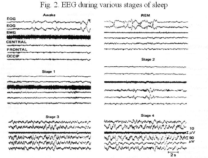
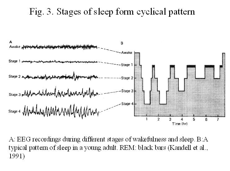
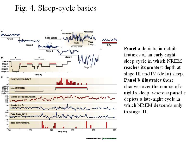
Sleep
Patterns. There are two different kinds of sleep: rapid eye movement (REM)
sleep and non-REM or slow-wave sleep. Non-REM sleep can be divided into several
stages (Figs. 2, 3, 4). A
person falling asleep first enters stage 1, which is characterized by
slight slowing of the EEG. Stage 2
is marked by the appearance of sleep spindles (12-14Hz) and high voltage
biphasic waves called K complexes, which occur episodically against a
background of continuing low voltage EEG activity. As sleep deepens, waves with
slower frequencies (0.1-4 Hz, mainly delta) and higher amplitude appear on the
EEG (Stage 3 and 4). The characteristic of deep sleep is a pattern of rhythmic
slow waves, indicating synchronization.
REM/Paradox
Sleep. The high-amplitude slow waves seen in the EEG during sleep are
sometimes replaced by rapid, low voltage, irregular EEG activity, which
resembles that seen in alert animals and humans (Figs. 2, 4).
However, sleep is not interrupted: indeed, the threshold for arousal by sensory
stimuli and by stimulation of the reticular formation (RF) is elevated. The
condition has been called paradoxical sleep. There are rapid, roving eye
movements during paradoxical sleep, and for that reason is also called REM
sleep. There are no such movements in slow-wave sleep, and consequently it is
often called non-REM sleep. Another characteristic of REM sleep is the
occurrence of large phasic potentials, occurring in groups of 3-5, that
originate in the pons and pass rapidly to the lateral geniculate body and
thence to the occipital cortex. For this reason, they are called
ponto-geniculo-occipital (PGO) spikes. There is a marked reduction in skeletal
muscle tone during REM sleep despite the rapid eye movements and PGO spikes.
The hypotonia is due to increased activity of the reticular inhibiting area in
the medulla, which brings about decreases in stretch and polysynaptic reflexes
by way of both pre- and postsynaptic inhibition. REM sleep is also characterized by dreaming episodes.
Mechanisms
of Arousal. Initial Studies (1935-1980)
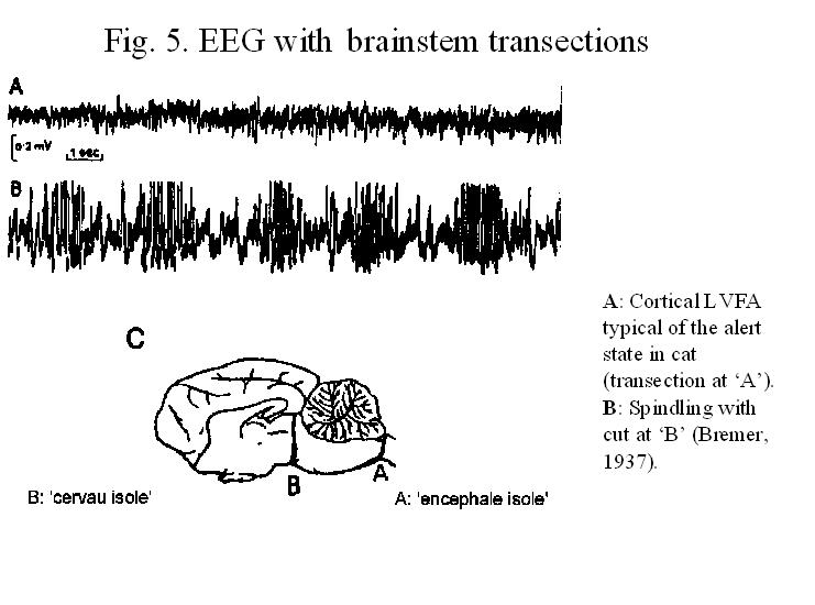
Bremer discovered in 1935 that when the
neuraxis of a cat is transected at Cl (encephale isole: Fig. 5.),
with artificial respiration and precaution for maintenance of blood pressure,
the animal shows the EEG and pupillary signs of normal sleep-wakefulness
cycles. In contrast, when the transection is made at the mesencephalic level,
just caudal to the motor nuclei of the third cranial nerve (cerveau isole:
Fig. 5),
there ensured a permanent condition resembling sleep.
Bremer's
discovery led to the concept of sleep as a passive process, as a deactivation phenomenon,
while, wakefulness is an active state maintained by afferent input to the brain
and sleep ensues when that input is removed, as in the cerveau isole
cat, or falls below a certain critical level, as in normal sleeping. In the cervau
isole preparation, olfactory input to the brain remains, but strong
olfactory stimuli produce only a transient activation that does not outlast the
stimulus. Visual pathways from the retina to the cortex are also intact, but
visual stimuli do not evoke widespread activation of the EEG in the cervau
isole animal, as they do in intact animals. Although Bremer tentatively
concluded that deafferentation per se is sufficient to induce sleep,
this last observation concerning visual stimuli indicates that some neural
mechanism in addition to the direct sensory pathways is required for the
maintenance of wakefulness.
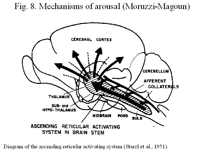
In
1949 Moruzzi and Magoun discovered that rapid stimulation (50-200/sec) of the
brainstem produced activation of the EEG (low voltage fast electrical activity,
or LFA), an effect evoked by stimulation of the central core of the brainstem
in a region extending upward from the bulbar RF to the mesodiencephalic
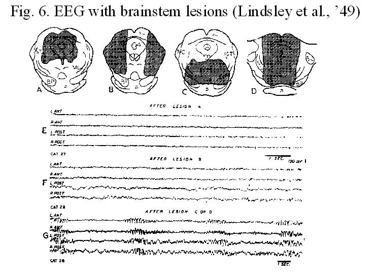
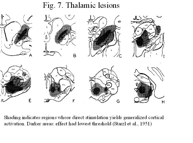
junction, the dorsal hypothalamus, and the ventral thalamus (Figs. 6, 7, 8). In
many features the activation produced by RF stimulation resembles the arousal
produced by natural stimulation. When the RF is stimulated via implanted
electrodes in sleeping animals, behavioral awakening and EEG desynchronization
result. This is also true in animals after section of the long ascending
sensory systems in the mesencephalon but does not occur after lesions of the
mesencephalic RF. Indeed, after extensive lesions of the mesencephalic RF,
animals may be comatose for many days and unresponsive to any stimuli (Lindsey
et al., 1949; French and Magoun, l952). If they survive, they may show good
recovery of sensory and motor functions but display various and sometimes
prolonged periods of somnolence, with marked refractoriness for arousal, which
when evokable, may not outlast the arousing stimuli. In contrast, animals
surviving transection of the long ascending and descending tracts of the
midbrain, but with no RF lesion, show no alterations of the sleep-wakefulness
cycle, are readily aroused and then show activated EEGs, although they are
profoundly deficient in the sensory spheres (Figs. 6).
Subsequently
by neuroanatomic techniques it was determined that the neurons of the RF
receive collateral input from visceral, somatic, and special sensory systems
and send long ascending projections into the forebrain via a dorsal pathway to
thalamic nuclei and a ventral pathway to and through the hypothalamus,
subthalamus and ventral thalamus and hence primarily through the intralaminar
thalamic nuclei to the cortex (Jones and Yang, 1985). The ascending reticular
system was thus identified located in the brainstem core and giving rise to
long ascending forebrain projections, that was necessary and sufficient for the tonic maintenance of the cortical
activation and behavioral arousal of wakefulness (Fig. 8). The possibility was considered that a
background of maintained activity within this ascending brain stem activating
system may account for wakefulness, while reduction of its activity either
naturally, by barbiturates or by experimental injury and disease, may
respectively precipitate normal sleep, contribute to anesthesia or produce
pathological somnolence.
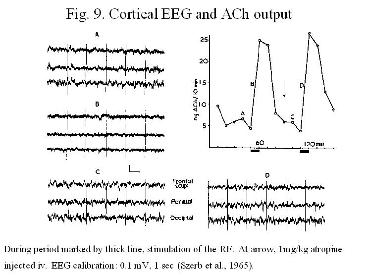
Later,
Szerb, Jasper and their coworkers showed (1965) that parallel to EEG
desynchronization during arousal or paradoxical sleep there is an increased
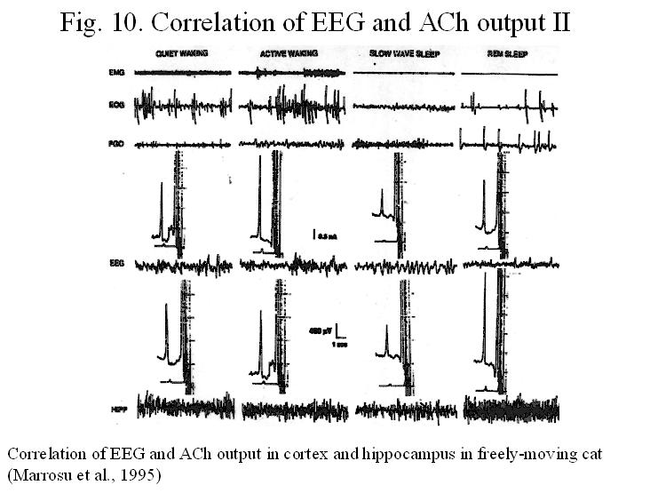
release of acetylcholine (ACh) over the whole cortex (Figs. 9).
The correlation of the different EEG epochs with the amount of ACh released in
the neocortex and hippocampus was confirmed recently using the more
sophisticated technique of in vivo dialysis (Marrosu et al., 1995). (Fig. 10.).
In
the 1920s, von Econonomo concluded that a “sleep regulating center” was present
within the midbrain and diencephalon.
Subsequent clinical studies (ref.:
Further investigations in the 1960s
and 1970s indicated that in the chronic course, the brainstem reticular
formation was not absolutely necessary for wakefulness, because cortical
activation could eventually recover, given sufficient time after lesions or
transections. Although ablation of the thalamus does lead to a temporary loss
of cortical activation; however, in the chronic course, cortical activation
does return. Furthermore, cortical
desynchronization can still be elicited by stimulation of the midbrain
reticular formation immediately after thalamic ablation, which indicates that
another, alternate extrathalamic route and relay to the cortex must exist. With
the development of increasingly sensitive biochemical, histochemical and
immunocytochemical techniques in combination with tracing studies confirmed the
presence of several extrathalamic corticopetal pathways that may participate in
regulating state related behavioral changes.
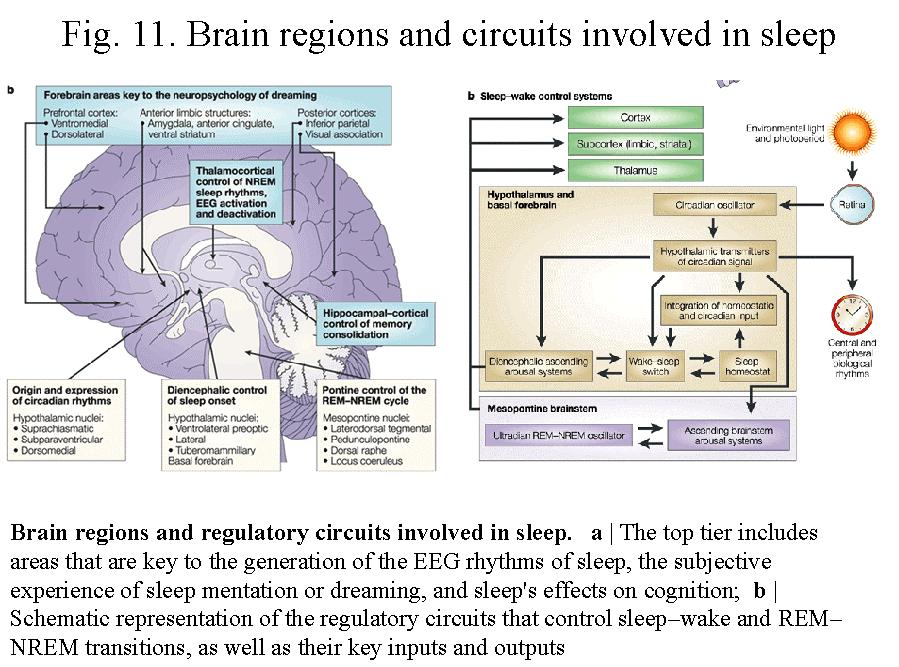
Figure 11.
summarizes brain regions and regulatory circuits involved in sleep. The
sleep-wake cycle is a complex phenomenon:
it is characterized by specific cortical EEG waveforms and synchronized
electrical activity (oscillations) in large scale networks, in particular in
the corticothalamic system. It is assumed that sleep-wake transitions are
accomplished by coordinated interactions between neural circuits of the
hypothalamic circadian, the mesopontine ultradian REM-non-REM oscillators and
GABAergic neurons of the ventrolateral preoptic area. Changing levels of
adenosine and other substances, acting via specific receptors in these circuits
mediate the homeostatic sleep pressure. The sleep-wake cycle is modulated by
activity of the brainstem, and forebrain arousal systems that use noradrenaline,
serotonin, histamin, acetylcholine and orexin/hypocretin among others as their
transmitters.
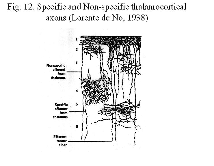
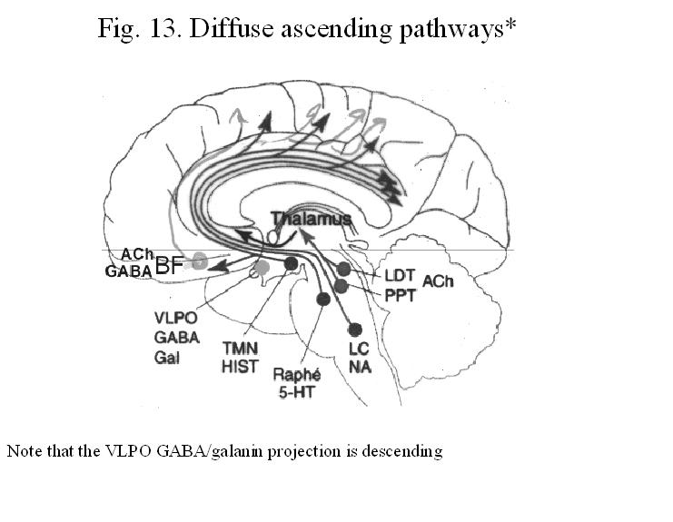
DIFFUSE ASCENDING MODULATORY SYSTEMS
Lorente de No (1938) noticed that two
types of fibers enter the cerebral cortx: one terminate primarily in layers III
and IV of a restricted area of the cortex, the second give off multiple radially oriented collaterals that innervate
primarily LI and VI over wide areas in the cortex (Fig. 12).
He called the first type of fibers ‘specific’, while the second ‘non-specific’.
He thought that specific fibers originate in the specific sensory thalamic
nuclei mediating visual, auditory and somatosensory information. On the other
hand, he thought that non-specific fibers originate in the so-called
non-specific (intralaminar, medial and midline) thalamic nuclei. Anatomical
studies in subsequent years established that the non-specific afferents to the
cortex originate in addition to the intarlaminar thalamic nuclei, in several
brainstem and forebrain regions and together represent the diffuse
extrathalamic corticopetal systems that will be described in detail below (Fig. 13)..
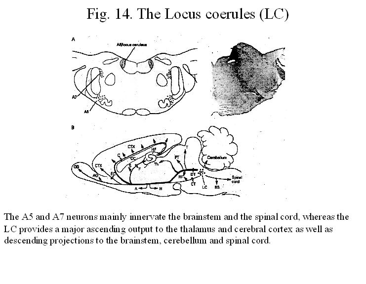
1. The Noradrenergic- Locus Coeruleus-Cortical
Projection (Figs. 14, 15, 16)
Anatomy. Considerable
evidence indicates that the locus coeruleus (LC) noradrenergic (NE) projection
to the cerebral cortex is highly collateralized, both within the cortex and between
it and other structures. There may also be a crude medial-to-lateral
topographical ordering to the coeruleocortical projection, but the
distributions of cells projecting to different cortical sites largely overlap.
Immunohistochemical studies, using an antibody against
dopamine-beta-hydroxylase (the enzyme noradrenaline) suggest that noradrenergic
axons establish conventional synapses in the cortex.
Noradrenergic
fibers in the cerebral cortex are densest in layer I (LI), where they are
mostly oriented parallel to the pial surface. Scattered fibers in LII and LIII
are mostly radially or tangentially oriented. The density of noradrenergic
fibers in LIV is greatest in granular primary sensory fields, in which the
axons are relatively short and run in an oblique direction.
Physiology.
Coeruleocortical neurons in rats and monkeys show long-duration action
potential and slow conduction velocities. Like mesolimbic cortical cells have
autoreceptors and apparently inhibit themselves by means of recurrent
collaterals acting on alfa-2 receptors, which cause hyperpolarization and a decrease
in membrane resistance. LC neurons tend to fire synchronously, often in bursts
in response to peripheral sensory stimuli; this is usually followed by a
quiescent period, which is thought to represent autoinhibition.
Studies on the effects of NE on
neurons in sensory cortical areas suggest that the net result of NE release is
an improvement in the signal-noise ratio. The noradrenergic innervation
together with the cholinergic one in the cerebral cortex plays an important
role in maintaining the plasticity of cortical connectivity (ocular dominance
shift in the visual cortex).
During wakefulness, the discharge
rates of LC neurons are closely tied to the state of arousal, as measured
electroencephalographically. During sleep, LC neurons in rats, cats and monkeys
show a progressive decrease in firing rate as slow-wave sleep deepens, then
become nearly silent before the onset of rapid eye movement or desynchronized
sleep. Neurons in the cerebral cortex,
thalamic reticular nucleus and thalamic relay nuclei change their activities in
vivo from periodic and rhythmic spike bursts during natural, slow wave
sleep to tonic firing of trains of single spikes during waking and REM sleep in
behaving cats with chronic implants. Similar changes in firing pattern occur in
vitro neurons in the cerebral cortex, thalamic reticular nucleus and
thalamic relay nuclei in response to NE. The slow depolarization results from
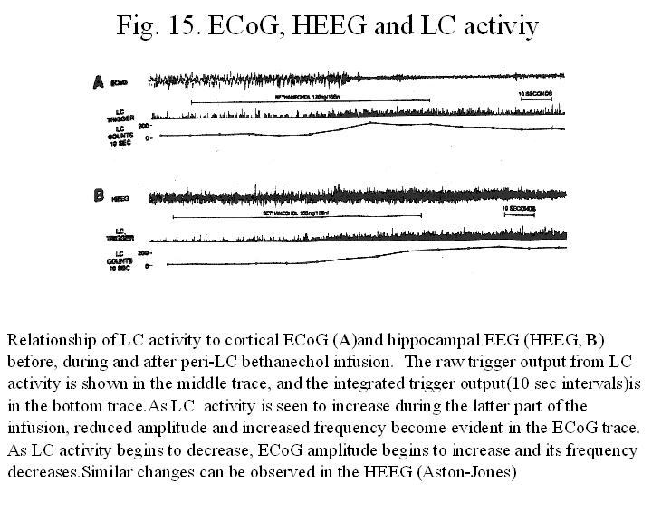
the reduction of K+ conductances and the enhancement of Ih. Peri-LC
bethanechol infusion results in an increase firing of LC neurons that is
followed consistently, within 5-30 sec, by a shift from low-frequncy, high
amplitude to high frequency, low amplitude activity in the neocortical EEG. The
infusion-induced changes in EEG are blocked by pretreatment (icv) with the
alpha-2 agonsit clonidine or beta-antagonist propanolol. Injection of clonidine
bilaterally immediately adjacent to LC induced a shift in neocortical EEG.
These observation indicate that the level of LC activity are not only
correlated with, but causally related to EEG measures of forebrain activation (Fig. 15).
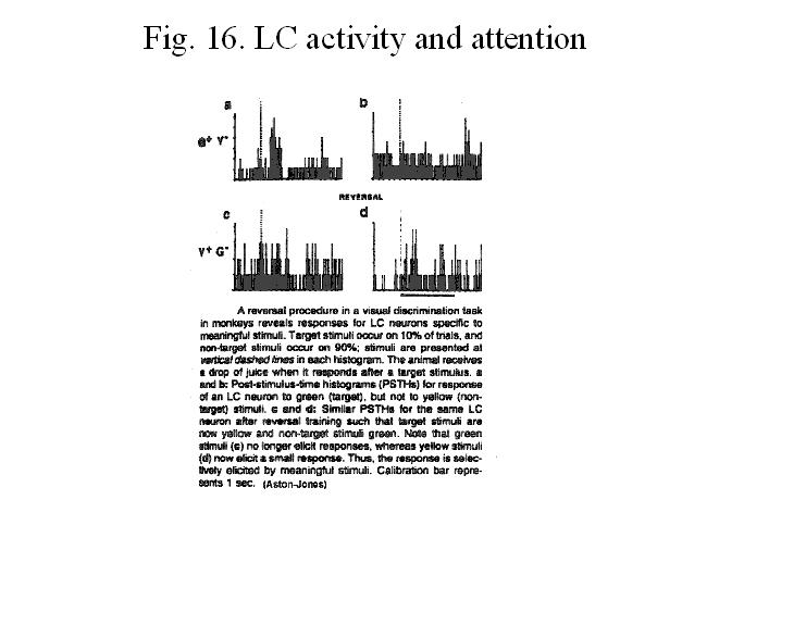
In addition to
changes in LC discharge preceeding corresponding changes in the EEG, LC
discharge rates also covary with orienting behavior. LC discharge associated
with orienting behavior is phasically most intense when automatic, tonic
behaviors (sleep, grooming or consumption) are suddenly disrupted and the
animal orients toward the external stimuli. Also, LC discharge closely
correlate with attentional behavior (Fig. 16).
Evidence also indicates that moderate LC ctivation accompanies optimal
information processing, whereas high discharge rates accompany, and perhaps,
produce a hyperarousal that may lead to poor performance in circumstances
requiring focused, sustained attention.
2.
Other Noradrenergic Cell groups in the Brainstem
A1 area. The A1 noradrenergic cell group area (Dahlström
and Fuxe, 1964) is located caudally within the ventrolateral medulla. Although
some fibers project to the midline thalamus, zona incerta and rostral
intralaminar thalamic nuclei, a considerable proportion of fibers ascend in the
medial forebrain bundle (MFB) and terminate in various hypothalamic nuclei, as
well as in basal forebrain areas containing
cholinergic projection neurons.
A2 area. The A2 noradrenergic
cell group is located in the caudal portion of the nucleus of the solitary
tract (NTS). It has been estimated that at least 90% of all nucleus
commissuralis (caudal, noradrenergic part of the NTS) neurons projecting
through the MFB are catecholaminergic (Moore and Guyenet, 1983). Projections
from this caudal part of the NTS in rat were followed to the lateral
parabrachial area, substantia innominata (SI), central amygdala and lateral bed
nucleus of the stria terminalis (BSt) (Norgren, l978; Ricardo and Koh, l978).
It is thus possible that basal forebrain cholinergic (BFC) cells receive general
viscerosensory input from the vagal nerve mediated through noradrenergic
afferents. On the other hand, different peptides such as enkephalin,
somatostatin, or substance P have been localized in ascending projections from
the caudal NTS (Riche et al., l990; Sawchenko et al., 1990), and are known to
be present in fibers within forebrain regions containing BFC neurons.
Therefore, it is possible that BFC neurons receive some peptidergic projections
from the NTS.
A5 area. The A5 noradrenergic cell group area
(Dahlström and Fuxe, l964) is located in the caudal pons, dorsolateral to the
superior olive. The projections of the A5 noradrenergic cell groups have been
described by Byrum and Guyenet (l987).
Noradrenergic neurons from this region project to several hypothalamic,
thalamic, and limbic nuclei, and it is possible that a subpopulation of BFC
neurons in the caudal SI receive such input. In view of the widespread
interconnections of the A5 group with basal forebrain regions involved in
cardiovascular regulation (Guyenet and Byrum, l985), it is possible that this
information also reaches the BFC system.
A7 area. The A7 noradrenergic cell group, which is
located between the ventrolateral border of the superior cerebellar peduncle
and the lateral lemniscus, constitutes a continuation of the A5 cell group.
Although in combined lesion-biochemical experiments it was reported that
ascending noradrenergic fibers from the area of the A7 cell group contribute to
the innervation of the hypothalamus (Palkovits et al., 1980), due to the fact
that fibers originating from more caudal noradrenergic cell groups project
through the A7 area, these results must be interpreted with caution. It is thus
not clear at present whether the A7 cell group projects to the basal forebrain.
3. Raphe-Cortical
Projection (Fig.
17)
Anatomy. The cortical serotoninergic innervation
arises in the dorsal (DR= dorsal raphe)
and superior central raphe nuclei, cell groups located ventral to the
cerebral aqueduct along the midline of the brainstem. Ascending fibers travel
primarily in a paramedian position trough the midbrain reticular formation and
ventral tegmental area (VTA) to the diencephalon, where they enter the MFB.
From this point, their course is similar to the other diffuse cortical
projection systems: a lateral systems of fibers turns laterally and runs
through the SI to external capsule, while a medial pathway continues rostrally
through the septum, dividing into a branch that runs back through the fornix to
the hippocampal formation and another branch that runs over the genu of the
corpus callosum and into the frontal cortex and cingulate bundle. The median
raphe nucleus contributes primarily to the medial pathway, whereas the dorsal
raphe fibers contribute to both projections.
Physiology. The electrophysiological
characteristics of serotoninergic neurons in the dorsal and median raphe nuclei
are in many ways similar to those of noradrenergic neurons. Specifically, raphe
neurons discharge at a relatively slow, regular rate, have long-duration action
potentials (3-4/ms), posses slowly conducting axons and show evidence of
inhibitory autoreceptors. Intracellular recording studies shows that the slow,
regular firing rates of dorsal raphe neurons is related to "pacemaker" potential in these
neurons. The activity of 5HT neurons in the dorsal and median raphe nuclei in
the unanesthetized cat relates closely to the wake-sleep cycle. During active
wakefulness the discharge rate averages 3.5 impulses/s. With the onset of
drowsiness, the rate begins to fall, and about 2-10 s before the onset of REM
sleep, the raphe neurons fall silent (Fig. 18).
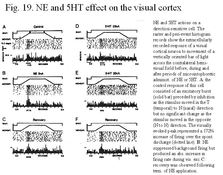
Iontophoretic application of 5HT to cortical neurons suggest that, like NE, the
effect of 5HT on cortical neurons may depend on the ongoing state of activity
of the target neuron (Fig. 19).
Electrical stimulation of the raphe is very effective in inducing neocortical
activation, this effect can be blocked by serotoninergic receptor anatgonits
such as ketanserin. Similarly, cortical activation induced by noxious
stimulation such as tail pinching, an effect that involves the 5HT systems, is
blocked by serotoninergic depletion (Dringenberg and Vanderwolf, 1998).
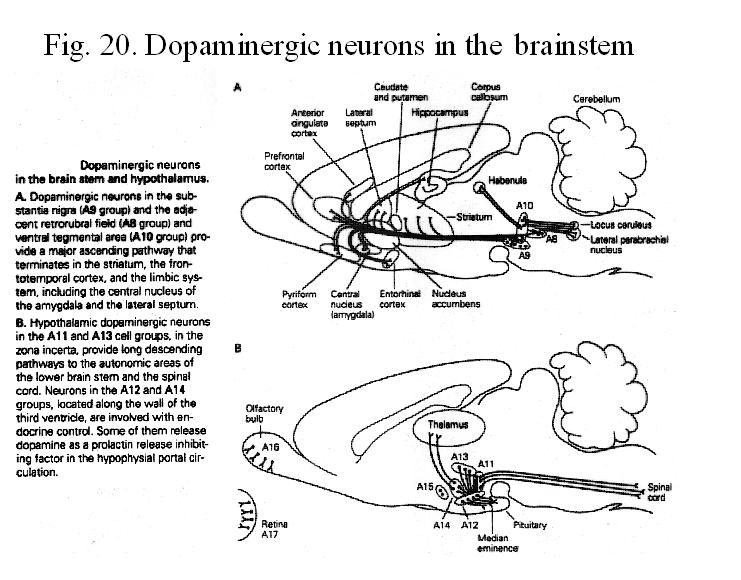
4. The Midbrain Dopaminergic System (Fig. 20)
DA neurons are concentrated in several cell groups in the brainstem. DA
neurons have homeostatic and regulatory roles n that they allow the forebrain
and cortical neuronal systems to function normally. A lesion of the midbrain
dopaminergic neurons disturbs many of the brain integrative functions not
directly related to sensory, motor processes or arousal. Lesion of the ventral
tegmental area (A10 or VTA) results in hypoactivity, a complete blockade of the
locomotor stimulating effect of amphetamine, aphagia, adipsia, deficit in
initiation and incentive to respond in an avoidance task, frontal neglect
syndrome, attentional impairments. Mesoaccumbens lesions cause an inability to
switch from one behavioral activity to another. DA neurons are activated when
the animal is presented with a behaviorally relevant stimulus requiring a
response. However, the DA system appears to be primarily involved during the
acquisition phase of this event, with little or no activation when the animal
is overtrained on the task (Schultz). According to Schultz the DA neurons
generate an error signal in the prediction of reward.
The firing rate or pattern of DA neurons
in the VTA and SNc is not significantly modulated by the sleep-wake cycle or
anesthetics. However, mice
with deleted dopamine transporter show increased wakefulness and decreased NREM
sleep. Furthermore, sleep disturbances in Parkonson’s disease and their
alleviation with dopaminergic medication suggest involvement of the
dopaminergic system in sleep-wake regulation (Aldrich, 2000).
5. Hypothalamocortical Projection
Hypothalamic lesions cause profound and
prolonged coma, which in monkeys or humans may last for years. These observations
suggest that the destruction of hypothalamic neurons that innervate the
cerebral cortex causes an irreversible deficit in cortical function.
Four distinct
hypothalamic cell groups that project to the cerebral cortex have been
distinguished.
1) In the tuberal
lateral hypothalamus, cortical projection neurons are located in clusters in
the zona incerta, the perifornical area, and along the medial edge of the
internal capsule. These neurons innervate the entire cortical mantle,
predominantly on the ipsilateral side. Many of the neurons in the perifornical
region contain orexin/hypocretin.
2) In the fields of
Forel at the premammillary level, a small group of neurons just ventral to the
medial tip of the medial lemniscus provides innervation primarily to the
ipsilateral frontal cortex.
3) Extending from the
posterior lateral hypothalamic area into the suprammillary nucleus is a dense
cluster of neurons that topographically innervate the entire cerebral cortex.
4) Neurons in the
tuberomammillary nucleus (TMN) on each side of the brain innervate the entire
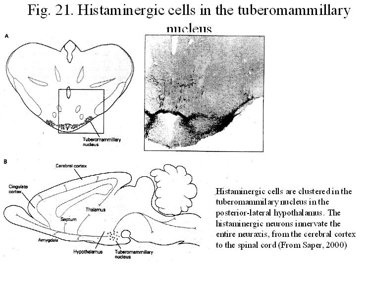
cerebral cortex bilaterally, many of these neurons synhetize histamin .
Histaminergic
(H) neurons (fig. 21). The histaminergic system innervates the
entire forebrain as well as brainstem regions that are involved in
behavioural-state control. A number of recent report suggest that histaminergic
projections from the tuberomammillary nucleus of the hypothalamus may act to
modulate EcoG actibvity and sleep-waking states. Intracerebral or
intraventricular administration of H or histaminergic agonists appears to
produce neocortical activation. However, like NE Histamine may not play a role
in the direct activation of the neocortex, and may produce its modulatory
effects on the EcoG by an indirect action via the cholinergic or serotoninergic
systems. After large depletion of brain histamine with
alfa-fluoromethylhistidine, both atropine-sensitive (cholinergic) and atropine-resistant
(serotonin) neocortical activation are intact, indicating that H is not
essential for the direct induction of cerebral activation. For example,
histamin microinjection into the basal forebrain-POAH area produce
dose-dependent increase in wake and blockade of histamine synthesis in the POAH
increases sleep and decrease wake.
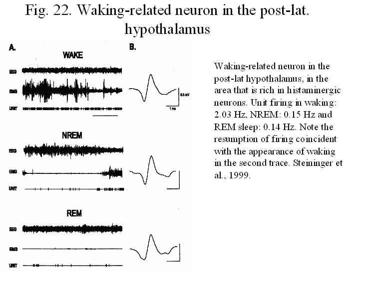
Neurons in this region in rats and cats,
using chronically implanted electrodes, were classified as waking-related (W),
W/REM-related and REM-related. W-related neurons decreased their discharge in
NREM sleep, and remained at low rates during REM sleep. A subpopulation of
these neurons disharge very little during REM sleep, and qualified as REM-off
neurons (Fig.
22). It is suggested that these latter units may correspond to
histaminergic neurons. Thus the histaminergic neurons fire in relation to the
EEG with a pattern similar to that of the noradrenergic and serotonergic
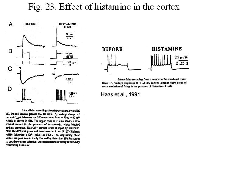
neurons of the lower brainstem. This is compatible with a action of histamine
on cortical neurons as reducing the accommodation of firing (Fig. 23).
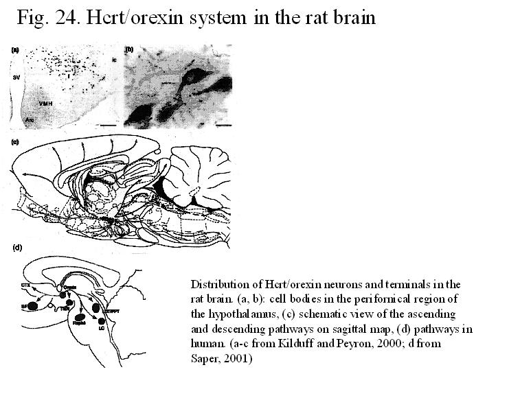
Orexin/hypocretin
(Fig. 24). Orexin cells are
localized exclusively in the tuberal region of the hypothalamus ventral to the
zona incerta and extend 1 mm rostrocaudally (in rat) behind the paraventricular
nucleus. In addition to food intake regulation, this system has been implicated
in neuroendocrine, cardiovascular, gastrointestinal control, water balance.
Mutation in the hypocretin receptor or the absence of ORX (hypocretin null mutant mice) cause in mice
periods of behavioral arrest that strongly resembled the cataplectic attacks
and sleep-onset REM periods characteristic of narcolepsy in dogs and humans.
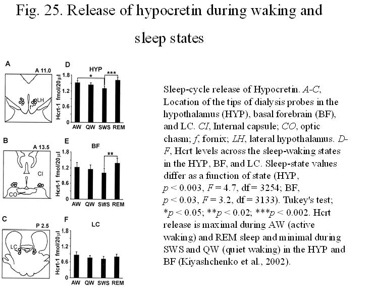
The release of orexin/hypocretin shows state-related changes: it is smaller in
SWS than quiete wake and REM sleep (Fig. 25)
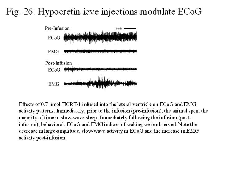
and ICV injections of ORX into rats at
light onset (the major sleep period) increases arousal and locomotor activity
and decreases REM sleep without affecting non-REM sleep (Fig. 26).
The effect of orexin in addition to its direct cortical projections is mediated
via the widespread projection of ORX
cells (Kilduff and Peyron, 2000). These
neurons project in addition to the neocortex to such diverse regions, as the
basal forebrain, preoptic area, TMN, DR, LC, mesopontine tegmentum, nuclei that
are all involved in behavioral state control. Hypocretins operate through Hcrt1
and Hcrt2 receptors that show differential distribution. For example, in the
basal forebrain, septum and the pontine reticular formation, neurons express
mostly, while in the LC, the predominant receptor is Hcrt1.
Fos
expression in orexin neurons correlates positively with the amount of
wakefulness and negatively with the amounts of non-REM and REM sleep. This
finding, together with studies that intraventricular or basal forebrain
injections of hypocretins produced increase in wakefulness, suggest that the
activation of hypothalamic hypocretin neurons may promote or contribute to the
maintenance of wakefulness. Excitatory effects of hypocretins on noradrenergic
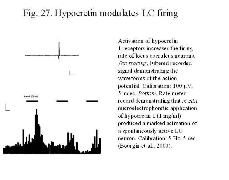
neurons of the LC (Fig. 27),
serotoninergic neurons of the dorsal raphe, histaminergic neurons of the TMN,
cholinergic neurons of the laterodorsal tegmental nucleus and cholinergic and
parvalbumin (PV) neurons of the basal forebrain have been described .
Hypocretin-containing axons establish asymmetric, excitatory type synapses on
septal cholinergic neurons and Hcrt2-receptors have been found on
parvalbumin-containing, septal GABAergic neurons .
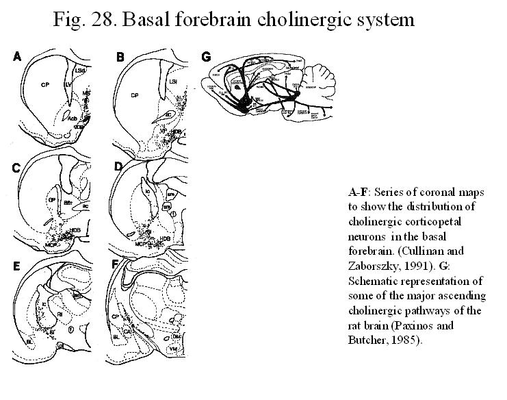
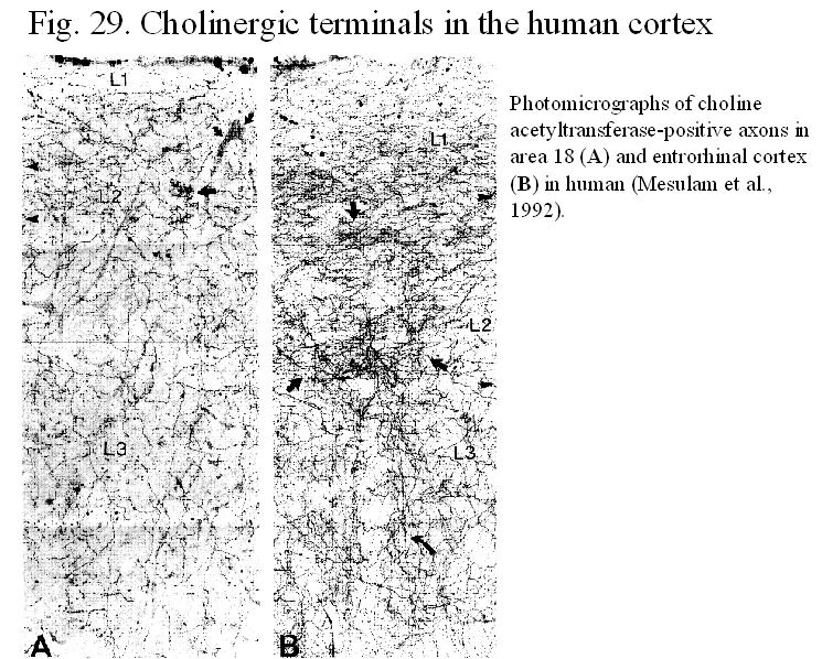
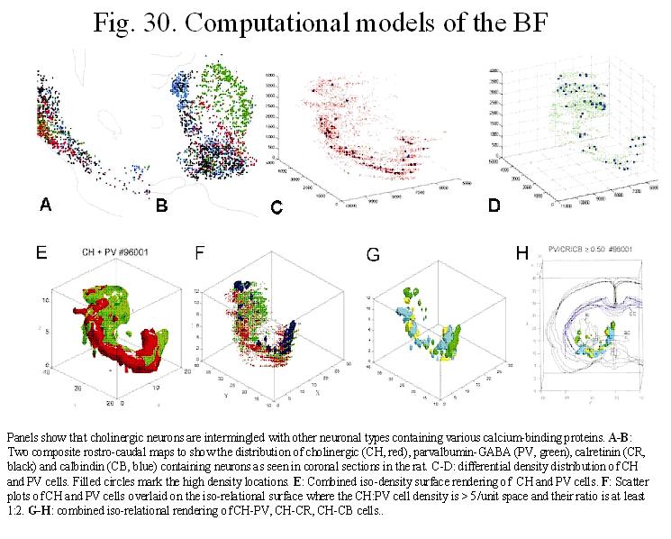
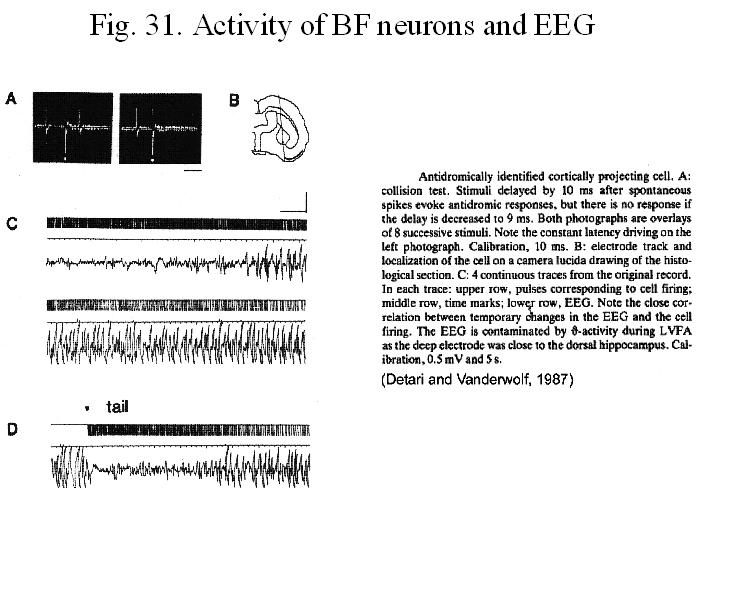
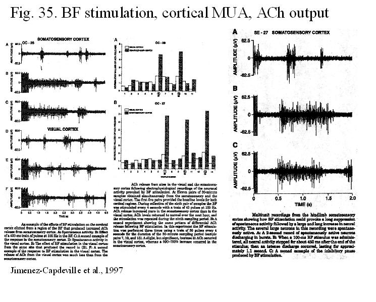
6. Basal Forebrain Corticopetal System (Figs. 28, 29, 30)
The magnocellular neurons in the basal
forebrain (termed in human Basal nucleus of Meynert) consists of a series of
clusters of large, darkly staining cortical projection neurons running through
several structures in the basal forebrain, including the medial septal and
diagonal band nuclei, the substantia innominata and peripallidal areas. In rat
cholinergic cells make up only about half of the neurons projecting to the
prefrontal and somatosensory areas, the rest is GABAergic or peptidergic.
GABAergic cells are often visualized using the presence of parvalbumin, a
calcium-binding protein in these neurons (Fig. 30).
The projection is topopgraphic and individual axons seem to innervate only
restricted cortical areas.
Basal
forebrain corticopetal neurons show rhythmic, spontaneous firing pattern (at an
average rate of approximately 20 impulses/sec) and the discharge rate of these
neurons is tightly coupled with cortical electrical activity: increased
discharge frequency of basal forebrain neurons during waking and REM sleep is
consistently associated with EEG desynchronization, while lower firing of BFC
neurons is paralleled with EEG synchronization. Electrical stimulation in the
basal forebrain results in short-latency excitation of neocortical neurons in
the frontal cortex, long lasting EEG desynchronization and release of ACh in
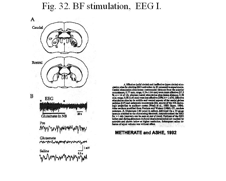
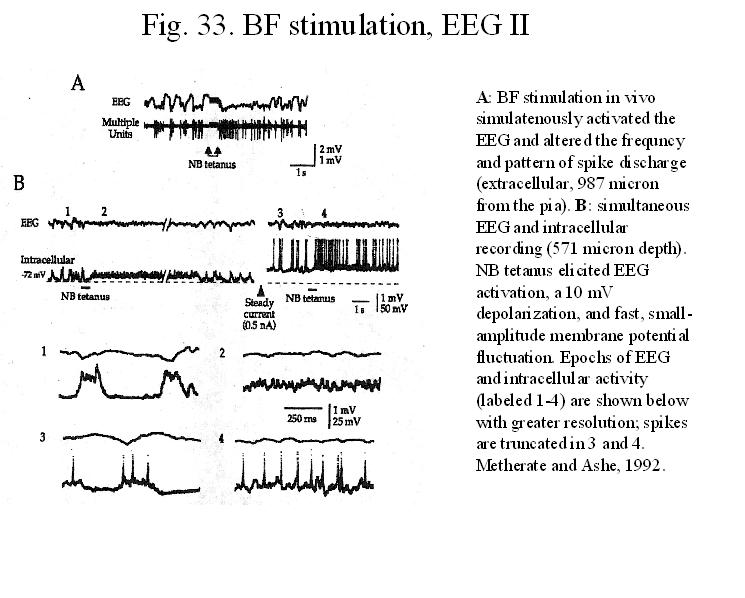
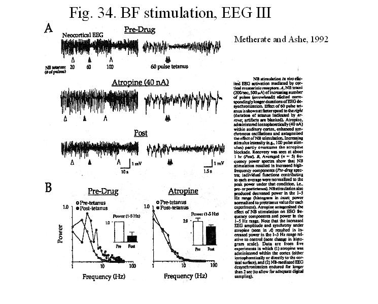
the cortex (Figs.31, 32, 33, 34, 35).
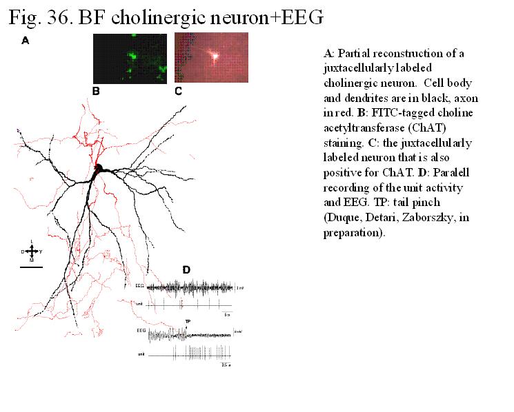
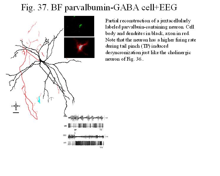
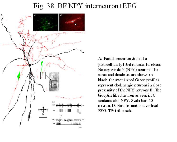
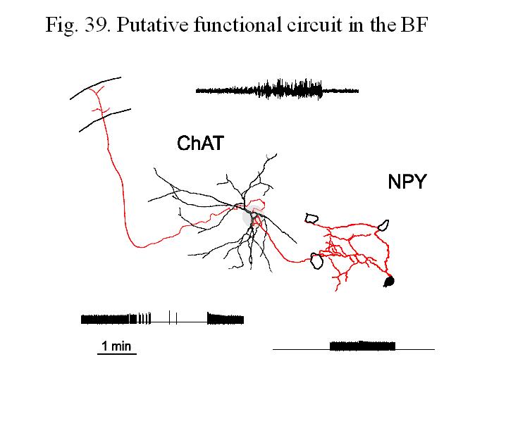
Recently, using the juxtacellular recording and filling technique, two types of
cortically projecting neurons (cholinergic, parvalbumin-containing GABAergic)
and a neuropeptide Y containing local neuronal type have been identified in
anesthesia while monitoring the EEG (Figs. 36, 37, 38).
Since the firing properties of cholinergic and NPY-containing neurons show opposite
pattern to the same EEG epoch, a possible functional circuit within the BF can
be envisaged (Fig. 39).
In
rats, the highest frequency activity of BF neurons was observed during running,
followed by drinking and immobility. The decrease in the firing rate correlated
with the increase of the power of slow activity in the neocortex. A further
decrease occurred in several BF neurons at the onset of high voltage
neocortical spindles, occasionally present during immobility in the rat. The
permissive action of BF neurons on spindle occurrence is also suggested by
increased incidence of spindling after damage to the BF and in aged rats with
shrunken cholinergic cells. These actions can also be explained by putative
inhibitory influences of basal forebrain cholinergic (BFC) neurons upon the
spindle pacemaker, on the reticular thalamic (RE) nucleus.
The mechanism, how in BFC neurons
low firing in slow wave sleep changes to more active state (in arousal or REM
sleep) is less well understood. It is likely that ascending noradrenergic
fibers from the locus coeruleus may
play an active role in alert state, while in REM sleep, when the locus
coeruleus and the raphe cells are silent, perhaps ascending glutamatergic axons
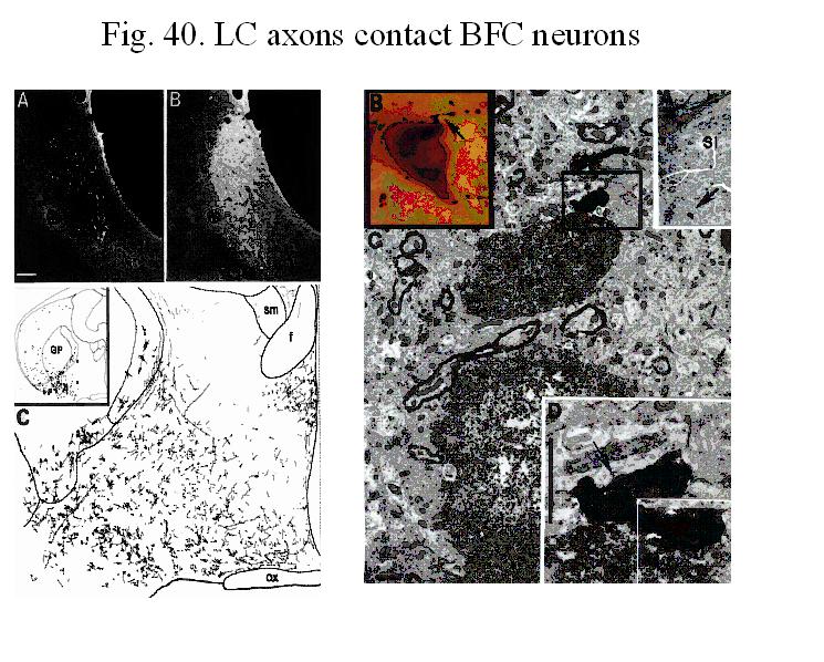
from the mesopontine tegmentum could stimulate BFC neurons. Indeed, LC axons
synapse on BF cholinergic neurons (Fig. 40) and
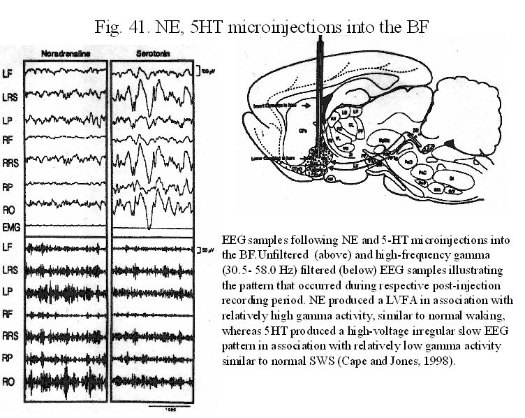
BF injection of NE affect specific cortical rhythm (Fig. 41).
Also, kainic acid injection into the substantia innominata of the basal
forebrain rapidly blocks the effect of reticular stimulation onto cortical
eveoked responses (Levandowski and Singer, 1993). In
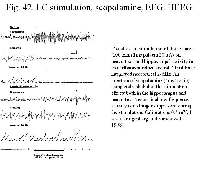
urethane-anesthetized rats stimulation of the LC area produces EcoG activation
in the neocortex and hippocampus and these effects are abolished by systemic
treatment with antimuscarinic drugs scopolamine or atropine (Fig. 42). These observation
suggest that the release of ACh and possibly the cholinergic input from the
basal forebrain (see below) to the cortex, play critical role in the EcoG
activation induced by the LC (Dringenberg and Vanderwolf, 1998).
During
arousal the BFC not only inhibit reticular thalamic bursting activity, but
through their projections in the neocortex, the released ACh in extensive areas
in the cortex provides a steady background of neocortical activity that may enhance
the effect of other afferents (for example those transmitting specific sensory
inputs) to the neocortex.
Summary of the diffuse
ascending modulatory systems
Although the original concept of Lorente de No about specific and nonspecific thalamocortical systems has not stood the test of time, nevertheless the diffuse cortical projection systems share, to a greater or lesser extent, certain anatomical and physiological features that make it useful to consider them as a whole. For example, in the rat all of the diffuse cortical projections tend to most heavily to innervate superficial layers (LI-II) and deep layers (LV-VI), avoiding the middle layers (III-IV) that in most areas receive the bulk of the specific thalamo-cortical projections.
Experiments involving
iontophoresis of monoamines or acetylcholine onto cortical neurons make it
clear that these substances primarily act, by means of complex effects on
membrane channels, to modulate the ongoing activity of the neuron. Instead of
serving strictly excitatory or inhibitory roles, these substances can either
enhance or impair discharge of the neuron to other inputs, and the total effect
depends on the physiological state of the target neuron.
Another emerging finding that supports this unitary view is the similarity of unit activity patterns in the various cortical projection cell groups. These observations suggest that the brainstem and basal forebrain projections to the cerebral cortex are primarily concerned with modulating the general level of cortical arousal as well as attention and motivation. The diffuse nature of this innervation, which includes the entire cortical mantle, and the prominent collateralization of the brainstem projections (particularly those from the locus coeruleus and raphe nuclei) are also consistent with a role in regulation of the overall level of cortical activity and mental state. On the other hand, the remarkable topographic specificity of the hypothalamic and basal forebrain projections to the cerebral cortex suggests that these diffuse cortical projections could selectively modify specific sensory, emotional or behavioral functions.
The Mesopontine Cholinergic Cells (Fig. 43). REM Sleep
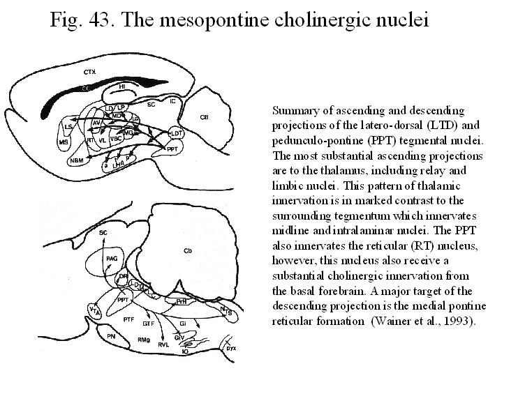
Figure 43. shows that most of the thalamic nuclei receive cholinergic input from two nuclei in the mesopontine tegmentum, the pedunculopontine (PPT) and laterodorsal tegmental (LDT) nuclei. The PPT and LDT in rat, monkey and human contain cholinergic and glutamatergic neurons whose axons project forward into the forebrain, particularly into the thalamic nuclei but also into the lateral hypothalamus and basal forebrain. A few cholinergic axon terminate in the prefrontal cortex. Lesions of the midbrain reticular formation, which diminish or eliminate cortical activation, as the early physiologists demonstrated, would destroy the cholinergic neurons located in the mesopontine tegmentum. However, more discrete neurotoxic lesions of the majority of the mesopontine cholinergic neurons did not produce any notable deficit in cortical activation (See Jones, 1994).
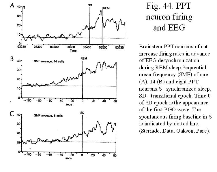
Neurons
in the brainstem PPT area shows increased firing in advance of EEG
desynchronization during REM sleep (Fig. 44).
That the brainstem reticular formation has a role in the blockade of
synchronized EEG rhythms is known from early experiments showing that periodic
spindle sequences appear on the EEG after transections at the collicular level
(Bremer’s cervau isole preparation) and that both spindles and slow waves
are readily erased by high-frequency
electrical stimulation of the upper brainstem
reticular core (Moruzzi and Magoun, 1949). Because passing fibers could
be activated by electrical stimulation, the use of microinjections of axon
sparing neurotoxins within the rostral brainstem reticular core helped to
demonstrate that perikarya in the rostral and caudal parts of the midbrain
reticular formation were indeed responsible for the EEG desynchronizing
reaction and behavioral arousal Since many components of these
brainstem-thalamic influences are antagonized by acetylcholine blockers, the
cholinergic projection from the mesopontine PPT and LDT nuclei were soon confirmed as important element in
the desynchronization process. Fig. 45-46 show
the location of cell groups involved in controlling the various events in REM
sleep.
REM Sleep
Paradoxical sleep (PS) was the term originally applied
by Jouvet and his colleagues in 1959 to periods of behavioral sleep during
which the eyes moved rapidly and the cerebral cortex showed a pattern of
activity similar to that of the waking brain in the cat. This unusual
association of parameters had been identified and described in humans several years earlier. This type of sleep
has according to its principal characteristics, been called PS, REM (rapid eye
movement) sleep, desynchronized sleep, active sleep, and dream sleep. The
principal and distinguishing characteristics of PS are low voltage fast
activity on the EEG, REMs recorded from the electrooculogram and muscle atonia
recorded from the neck electromyogram (EMG). During PS, the REMs are
accompanied by phasic activity within the visual system (PGO spikes). The
manifestation of this same phasic activity occurs peripherally as REMs and also
as twiches of facial, hypoglossal and distal limb muscles. At the same time
that this phasic activity is being internally generated, somatic reflexes are
inhibited, reflecting an inhibition of both sensory input and motor output.
Sensory transmission is inhibited by both presynaptic inhibition of the primary afferent fibers and
postsynaptic inhibition of sensory relay neurons. Somatic motoneurons of the
spinal cord and brainstem are tonically inhibited as evident by
hypewrpolarization of the membrane of these cells. Within the autonomic nervous
system, reflexes are also attenuated, as manifest by marked alteration of
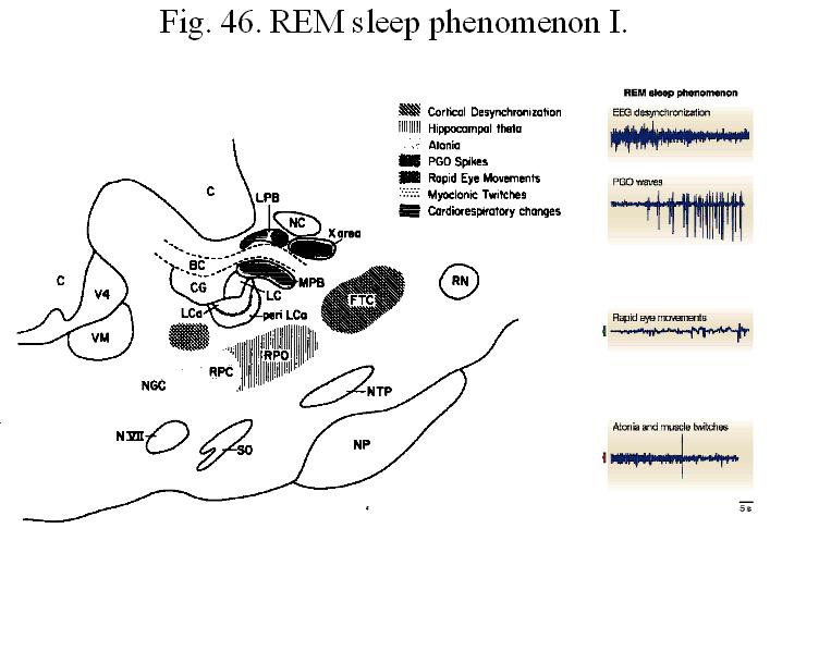
cardiovascular, respiratory and temperature regulation during this state. Fig. 45
summarizes the location of cell groups involved in controlling the major events in REM sleep.
PS occurs in a cyclic manner following a given period of
SWS which corresponds in the human approximately 45-85 min (progressing from
longer to shorter periods through the night). PS endures on the average 5min in
the cat and 5-65 min in the human. The average length of the sleep cycle
beginning with SWS and ending with REM sleep is approximately 90 min in man and
corresponds to a basic rest-activity cycle. This ultradian rhythm is normally
correlated with an ultradian tempertaure cycle
of approximately 90 min. Over the course of this cycle during sleep,
body and brain temperature decrease during SWS relative to waking and increase
during PS relative to SWS. In correlation with the cyclic temperature changes,
CBF and metabolism also change during the sleep cycle. Glucose metabolism is
also reduced during SWS and is increased to its highest levels through PS. Thus
the sleep cycle corresponds to a basic rest-activity cycle of the brain.
PS has been identified in most mammals and in birds.
Across mammals, the duration of PS is a
function of the sleep cycle length, that increases with the size of the body
and brain across species. PS has been consistently found to occur in its
greatest amounts in the fetus or immature newborn animal. This would suggest that PS may be crucial to
the development of functional circuits, such as those for co-ordinated eye-head
movements, locomotion or complex species-specific behaviors. Absolute and
prolonged deprivation of PS, like that of total sleep, leads to the death of
the animal associated with weight loss,
hypothermia in a period of two to eight weeks in adult rat. Thus PS can
be viewed as an important function both during development and in adulthood,
important perhaps for sensorimotor programming in development and information
processing though life and also more fundamentally vital for physiological and
metabolic functions of the brain not yet fully understood but as part of a
basic rest-activitry cycle.
The results of the transsections studies indicated the
importance of the pontine tegmentum in the generation of the phasic and tonic
activation as well as the inhibitory processes of PS. Transmission of phasic activation evident as PGO spikes from the
pons to the lateral geniculate, occurs along a pathway ascending from and
though the dorsolateral pontomesencephalic tegmentum. Tonic cortical activation, associated with the state of PS depend
upon multiple systems that relay activation from the branistem reticular
formation to the cerebral cortex and which in addition to the thalamocortical
relay, include a ventral, extrathalamic pathway and relay through the
hypothalamus and basal forebrain. Transections studies also suggested that the
medullary ventral reticular formation serves s the relay and final common
pathway in producing the inhibition within the spinal cord.
Following neurotoxic lesions of the pontomesencephalic
area, including the cholinergic neurons, PS was eliminated 2-3 weeks. Incipient
PS episodes reappered following 3 weeks and were characterized by low voltage fast EEG activity in
association with minimal PGO spike-like activity and minimal REM and in
association with abnormal persistence of neck muscle tone. These results
suggets that cholinergic neurons of the dorsolateral pontomes. tegmentum may be
critically invoplved in the initiation and maintenace of the state of PS and
the associated phasic PGO spikes. Pontomesencephalic cholinergic neurons have
been found to give rise long projections
into the forebrain, predominantllty to the thalamus and could thus
mediate a cholinergic influence upon EEG and PGO. Although cholinergic neurons
of the PPT/LDT area send descending projections through the tegmentoreticular
tract to the medullary reticular formation (RF), pharmacological studies do not
support a direct cholinergic role in the motor inhibition of PS. Specifically,
it seems that pontine tegmental neurons that receive a cholinergic innervation
may in turn via projections to the medullary reticular formation transmit
signals involved in the motor inhibition that naturally occcurs during PS. The
non-cholinergic neurons of the tegmentoreticular system may utilize glutamate
as transmitter, since injection of Glu into the medullary RF produce muscle
atonia. Neurons of the medial medullary RF may either relay or contribute to
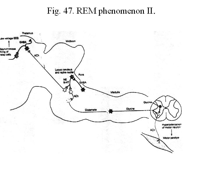
the reticulospinal influence that results in motor inhibition (Fig. 46).
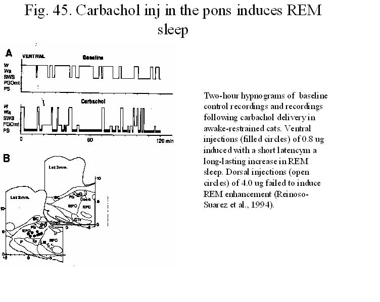
Carbachol, a cholinergic agonist injected into the ventral pontin oral nucleus
induce a long-lasting increase in REM sleep (Fig. 47).
Since cholinergic (REM-on) neurons are active during
PS while LC noradrenergic and serotoninergic raphe (REM-off neurons) cells are
silent there is a reason to believe that a direct or indirect interaction
between the cholinergic and monoaminergic system may underlie the fundamental
properties and generations of this state, as suggested by McCarley and Hobson
in the late seventies. In narcoleptic* dogs, biochemical studies have revealed
higher concentration of muscarinic agonists and the symptoms can be reduced by
muscarinic antagonists. Reciprocally, evidence indicates that both
catecholamines and 5-HT metabolism may be deficient and that drugs which
enhances synaptic concentration of monoamines can reduce the cataleptic or
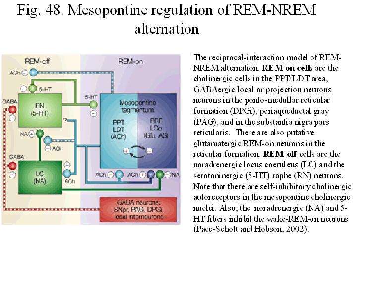
narcoleptic attacks in dogs and humans. Figure 48
summarizes the updated version of the reciproc-interaction model to explain th
eREM-nonREM alternation. As this scheme shows in addition to cholinergic cells,
REM-on neurons contain gluatamate and local GABAergic neurons. Additionally,
descending GABAergic projections from the preoptic area, ventral periaqueductal
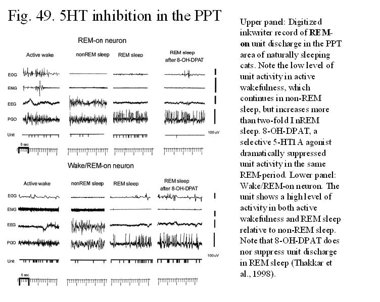
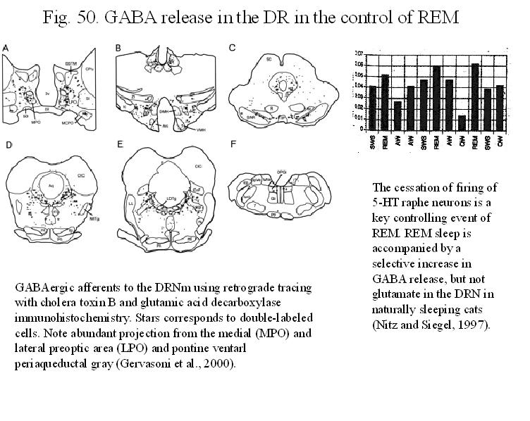
region contribute to the increased GABA release ( Figs. 49-50) during
REM sleep in the noradrenergic and serotoninergic nuclei.
___________________________________________________________________________________
*
Narcolepsy is irresistable
attacks of sleep associated with cataplexy, paralysis and/or hallucinations.
These attacks represent a sudden onset of REM sleep, motor inhibition and dream
activity.
-----------------------------------------------------------------------------
Thalamocortical
Oscillations in the Sleeping and Aroused Brain
Since 1980, major progress has been made in investigating the mechanisms of generating rhythmic activity in thalamocortical systems. Studies, using simultaneous intra and extracellular recordings in multiple sites of thalamic and neocortical areas both in vivo and in vitro as well as computer simulations have revealed the ionic conductances that contribute to the intrinsic oscillatory properties of neurons and also demonstrated how these oscillations of isolated neurons can be transformed by interactions with other neurons into rhythmic patterns (Steriade, McCormick, Sejnowski).
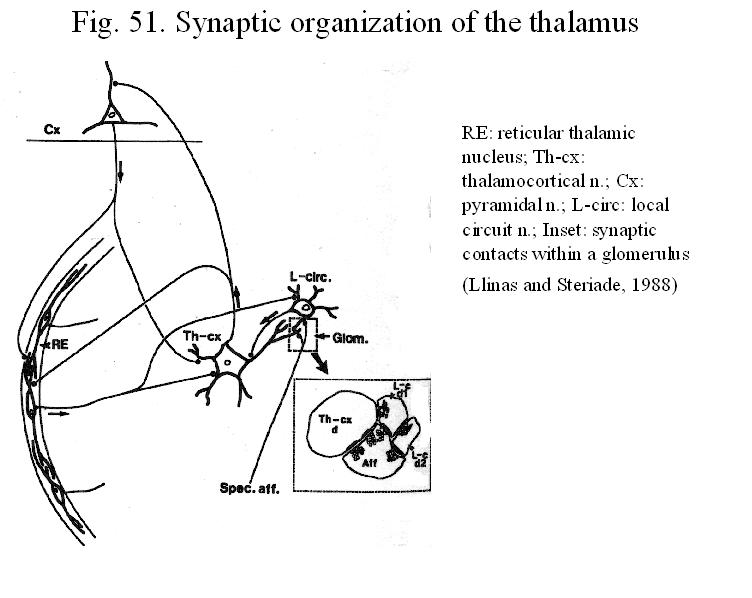
Fig. 51 shows the synaptic organization in the thalamus. Different areas of the cerebral cortex receive inputs from various thalamic nuclei. In turn, cortical neurons of layer 6 innervate topographically appropriate regions of both the dorsal thalamus and reticular nucleus (RE). The RE cells receive excitatory inputs from axon collaterals of thalamic neurons that project to the cortex and of cortical neurons that project to the thalamus; RE cells project to specific relay neurons and also innervate other cells of the RE. All neurons in the RE are GABAergic.
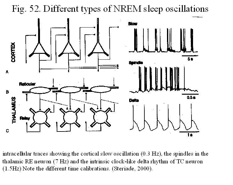
The majority of neurons in the mammalian brain have two basic modes of operation: tonic (steady) firing during EEG-desynchronized behavioral states and burst discharges during EEG synchronized sleep. The burst discharge mode appears to be an intrinsic features of several neuronal types. An extreme example of the complex interplay of sequentially linked ionic conductances is the oscillatory mode.
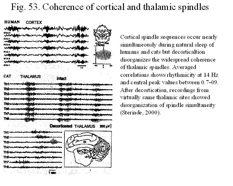
Figure 52 summarizes the different types of NREM sleep oscillations in the thalamo-cortical networks. Sleep spindles (with a frequency of 7-14 Hz=alpha waves) are the epitome of EEG synchronization during light sleep. Slow waves or delta waves (1-4 Hz), and slow rhythm (0.1-Hz) prevail during the deep stage of non-REM sleep. Figure 53. shows that cortical spindle sequences occur nearly simulatenously during natural sleep in humans and cats and decortication disrupt the widespread coherence of thalamic spindles. Three factors account for the appearance of spindle and delta rhythms. Two of them consist of intrinsic properties and ionic conductances that allow thalamic cells to oscillate and synchronizing synaptic networks that include the reticular thalamic nucleus. The other factor is the dampened activity in ascending cholinergic brainstem reticular projections that normally act to prevent the occurrence of, or to block ongoing spindle and delta oscillations.
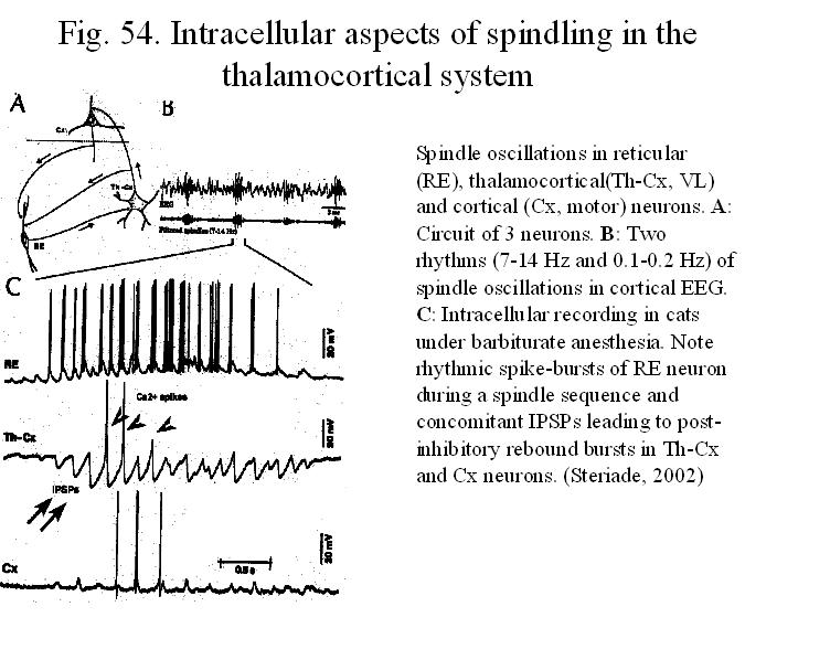
Spindle oscillations consist of waxing-and-waning field potentials of 7-14 Hz, grouped in
sequences that last for 1-3 s and recur once every 3 to 10 sec (Fig. 54).
The EEG spindles are the electrographic landmarks for the transition from
waking to sleep that is associated with loss of perceptual awareness. These
oscillations are generated in the thalamus as the result of synaptic
interactions in a network in which the main players are the inhibitory neurons
of the reticular thalamic (RE) neurons, thalamocortical relay cells and
cortical pyramidal neurons. Through their connections, the RE is uniquely
positioned to influence of the flow of information between the thalamus and
cerebral cortex. Intracellularly (Fig. 54),
spindles are characterized in RE neurons by a slowing, growing and decaying depolarizing
envelope with superimposed spike barrages, whereas in thalamocortical
neurons spindles are associated with cyclic long-lasting hyperpolarizations
that eventually lead to rebound bursts transferred to the cortical pyramidal
neurons. The synchronization of this oscillation between neighbouring cells in
either the RE or relay nuclei results from a large overlap in the afferent and
efferent connections. That the RE nucleus is the pacemaker of spindle
rhytmicity is demonstrated by abolition of spindle waves in RE deprived
thalamocortical neurons and preservation of spindle rhythms in RE neurons
disconnected from their thalamic and cortical inputs.
Delta waves.
High-amplitude, slow delta waves (1-4Hz) are most frequently observed during
stage 4 sleep in the normal brain. The rhythmicity of the cortical delta waves
is explained by the triggering effect of the periodic quasi synchronous
thalamocortical inputs. The thalamus can maintain a rhythmic oscillation in the
delta range due to hyperpolarization-dependent intrinsic property of
thalamocortical neurons and their network connectivity with the GABAergic
reticular nucleus. Delta waves occur with largest amplitude in deep layer V
cortical layers, and they are recorded as negative waves on the neocortical
surface or on the scalp. Depth profile measurements in the neocortex of the
cat, rabbit and rat revealed that surface-negative-deep positive delta waves during
SWS correlate with the suppression or cessation of discharges of
Although the intrinsic properties of thalamic neurons are fundamental in allowing them to oscillate, in intact brain, these properties are subject to controlling influences from modulatory ascending systems (cholinergic, noradrenergic, serotoninergic, histaminergic) that change the functional mode of single neurons as well as to the influence of a pacemaker (the reticular thalamic nucleus), which, by virtue of its connections to all thalamic nuclei, synchronizes the activity of thalamic neurons. The ascending modulatory axons collectively innervate the entire expanse of the cerebral cortex and the thalamus (both the relay and reticular nuclei). Through specific receptors, these transmitters induce changes in the membrane properties of the thalamic and cortical neurons promoting more tonic activity and inhibiting those ionic conductances which are responsible for the oscillatory mode.
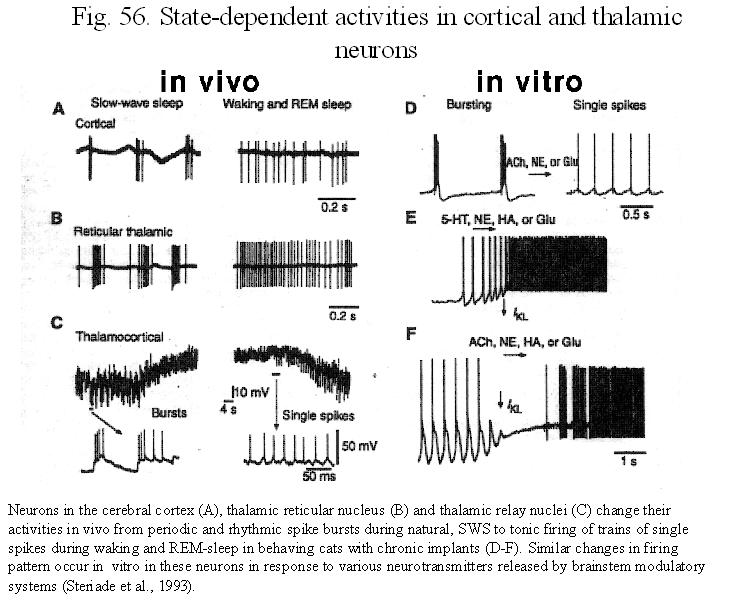
EEG desynchronization is characterized by the disruption of spindle oscillations in the thalamocortical systems during both waking and REM sleep and upon midbrain reticular stimulation (Fig. 55). The effects of the putative neurotransmitters released by ascending activating systems, as revealed by in vivo and in vitro experiments, confirm that all these neurotransmitters help maintain the waking state and for ACh, also the dreaming state (Fig. 56). The changes in firing between sleep and arousal are accomplished by depolarization of the membrane potential in the thalamocortical neurons by 5-20 mV, which inactivates the low-threshold Ca2+ current and therefore inhibit burst firing. Brainstem peribrachial stimulation blocks an ongoing spindle sequence in RE neurons by producing a large hyperpolarization (Fig. 55) associated with an increase in membrane conductance. Electrical stimulation in the region of brainstem cholinergic and noradrenergic neurons, or direct application of ACh or NE, results in prolonged depolarization of thalamocortical cells. In thalamocortical cells, these transmitter-induced depolarizations results from muscarinic ACh and alpha1 adrenergic receptors. The peribrachial-evoked hyperpolarization in RE neurons is a muscarinic effect, as it is blocked by scopolamine. The firing rates of neurons in brainstem PPT neurons increase in anticipation of awakening or before REM sleep (Fig.44) in further support of the origin of desynchronization.
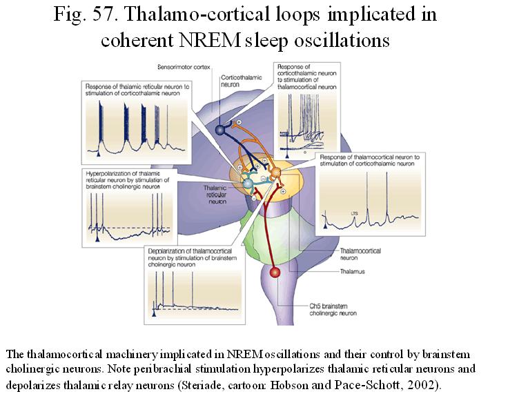
During wakefulness, enhanced synaptic excitability of thalamocortical systems is accompanied by an increased efficacy of the fine inhibitory sculpturing of afferent information. It has been already mentioned that brainstem modulatory systems, particularly the cholinergic one inhibits the spindles at their site of genesis, the reticular thalamic nucleus, ACh, however, also induces an enhancement of the stimulus-specific inhibition by excitation of local circuit neurons in the thalamic relay cells. The facilitatory effects of brainstem reticular stimulation and natural arousal on short-range specific inhibition has also been observed in corticofugal neurons during wakefulness (Steriade, 1994). Figure 57 summarizes in a cartoon the thalamocortical machinery in NREM oscillations and their disruption by the brainstem cholinergic neurons.
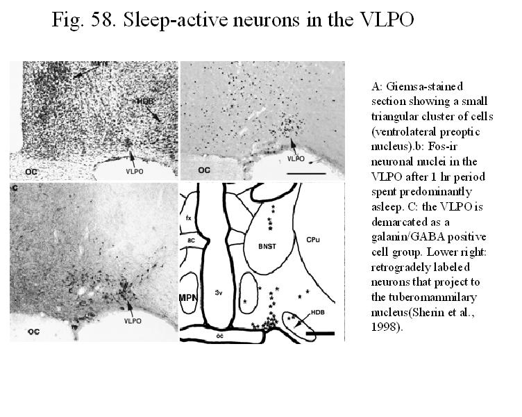
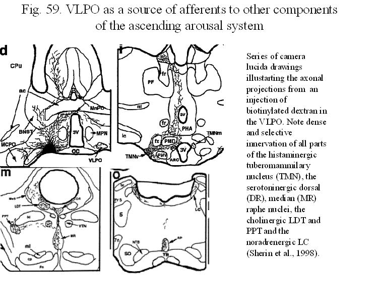
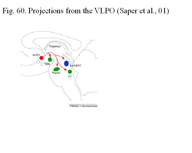
The
switch-off: sleep active neurons in the ventrolateral preoptic nucleus (VLPO)
A large body of
evidence suggest that neurons in the preoptic/hypothalamic area, adjacent to
the basal forebrain, play an important role in triggering sleep, especially
NREM sleep. For example, lesions
involving this region in humans, cats and rats induce long-lasting insomnia,
whereas its stimulation can be sleep-promoting in animals Furthermore, several
groups described cells in the preoptic/anterior hypothalamic areas
of cats and rats that increased their discharge in anticipation of non-REM
sleep onset
More recently, it has been shown that a dense
cell cluster in the ventrolateral preoptic area (VLPO) shows c-fos activation
proportional to the amount of time spent in sleep but not circadian time.
Moreover, the majority of these VLPO cells show elevated discharge rates in
both SWS and REM sleep as compared to waking. These neurons express
GABA/galanin and project to the hypothalamic tuberomammilllary nucleus.
Furthermore, a projection from the VLPO and the surrounding preoptic cells to
the locus coeruleus, dorsal raphe and PPT-LDT cell groups has been described.
It is suggested that this descending GABAergic pathway might promote REM sleep
by inhibiting the discharge of brainstem aminergic and cholinergic nuclei. Figs. 58, 59, 60 show
the location and projections from the VLPO.
Homeostatic
and Circadian Regulation of Sleep
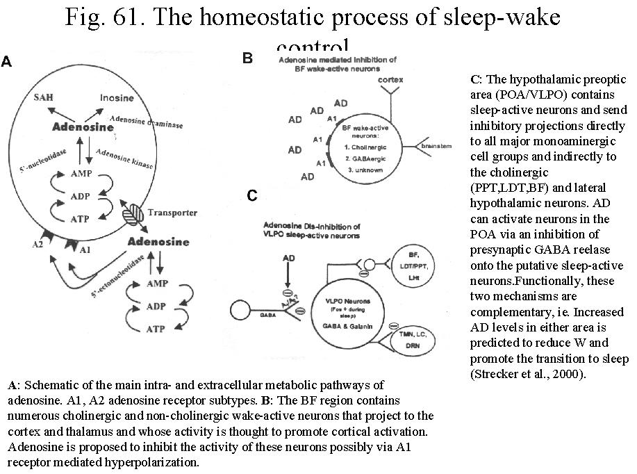
Recent
studies suggest that mesopontine and BF cholinergic neurons are under the tonic
inhibitory control of endogeneous adenosine, a neuromodulator released during brain metabolism.
Increased metabolic activity during
waking may cause an increase in both intra and extracellular adenosine.
Consequently, cholinergic neurons are under increasing inhibitory influence
through adenosine receptors. During the reduced metabolic activity of sleep, on
the other hand cholinergic neurons are slowly released from the adensoine
inhibition due to their low level of production. These suggestive data would
constitute a long sought coupling mechanism that links neuronal control of EEG
arousal to the effect of prior wakefulness (Strecker et al., 2000; Fig. 61).
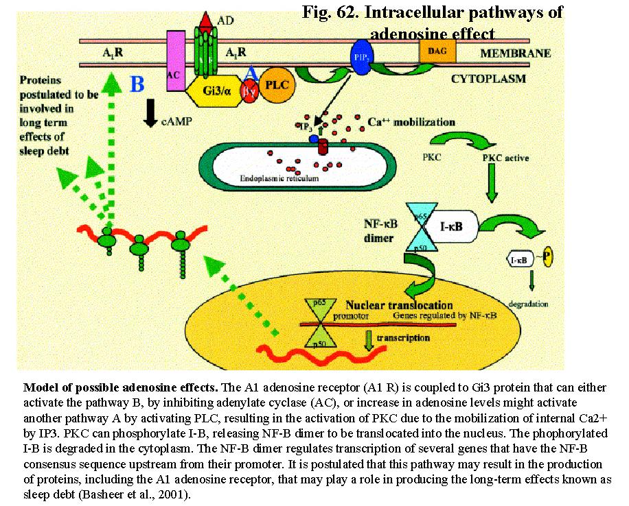
Binding of adenosine to A1 receptors in a subpopulation of cholinergic neurons
in the ventrolateral basal forebrain may preferentially activate the PLC
pathway to mobilize internal calcium that activate PKC. Activated PKC then
increases the DNA binding activity of the transcription factor, nuclear factor B (NF-
B) which is known to alter the
expression of several behavioral state regulatory factors, including
interleukin-1Beta, tumor necrosis factor-Alpha, nitric oxide synthase, cyclooxygenase-2 and even A1 adenosine receptor
mRNA. These changes may contribute to the long-term effects of sleep
deprivation (Fig. 62).for
review see Basheer et al., 2002).
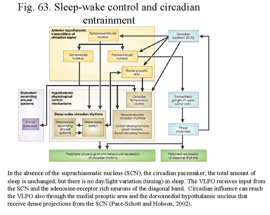
According to the two-process model of sleep
regulation (Borbely, 2001), the homeostatic sleep pressure with duration of
wakefulness must be integrated with circadian propensity to initiate sleep. In
the absence of the suprachiasmatic nucleus (SCN), the circadian pacemaker, the
total amount of sleep is unchanged, but there is no day/light variation in
sleep timing. The VLPO receives input from the SCN and retina and receive input
from adenosine receptor rich neurons of the diagonal band. Thus the VLPO is
anatomically well-positioned to integrate homeostatic and ciracadian drives and
to influence forebrain and brainstem arousal systems. Circadian influence can
reach the VLPO also through the medial preoptic area and the dorsomedial
hypothalamic nucleui that receive dense
projections from the SCN and projects to the VLPO (Fig. 63).
Summary of Sleep-Wake Cycle and Sensory Processing
1) Single unit recordings showed that the discharge
rate of thalamo-cortical and corticofugal neurons is generally higher in REM
sleep than in the waking state. In addition, the otho- and/or antidromic
excitability of these cells was the same or higher in REM than in awake
animals.
2)At the cortical level, evoked potential studies of
thalamic and cortical regions in different sensory modalities suggests that
their synaptic excitability diminishes from waking to SWS but surpasses waking values in REM sleep.
Finally, in contrast to wakefulness, REM sleep was accompanied by a reduction
of inhibitory activity in cortical neurons.
3) Studies in humans found that the percentage of
awakenings evoked by sensory stimuli decreased from stage I to stage IV with
REM sleep displaying intermediary values.
These studies draw our attention to the central paradox
of REM sleep. Namely, that stimuli which are perceived in the waking state do
not awaken subjects in REM sleep, even though the amplitude of the primary
evoked cortical responses is generally similar to or higher than, in the waking
state. In other words, although the thalamo-cortical network appears to be at
least as excitable during REM sleep as in waking state, the input is mostly
ignored. The lack of behavioral response to suprathreshold sensory stimuli
reflect a difference in the way the brain processes sensory input. REM sleep
can be considered as a modified attentive state in which attention is tuned
away from the sensory input toward memories.
The synaptic transmission of sensory information
through the thalamus and the cerebral cortex is enhanced during the states of
waking and REM sleep, compared with EEG-synchronized sleep. The
obliteration of synaptic transmission occurs in the thalamus at the first EEG
signs of drowsiness, before overt behavioral manifestation of sleep and despite
the unchanged magnitude of the incoming (prethalamic) volley. The amplitude of
the monosynaptically evoked wave of thalamic and cortical field response is
greatly increased both during EEG-desynchronized behavior states (waking and REM sleep) in chronic
experiments and on brainstem reticular stimulation in acutely prepared animals.
These changes are observed in all sensory and motor thalamocortical systems.
The synaptically relayed component progressively diminishes in amplitude from
the very onset of EEG synchronization during drowsiness and is completely
obliterated during EEG-synchronized sleep, in spite of the unchanged amplitude
of the presynaptic component. The blockade of synaptic transmission through the
thalamus prevents the cerebral cortex from elaborating a response and is a
necessary deafferentation prelude for falling asleep. Neocortical delta waves
indicates that the principal neurons of the cortex are engaged in a collective
burst mode of operation (synchronous hyperpolarization, synchronization), and the
EEG waves themselves reflect the long-lasting AHPs that follow such bursts.
This ‘closed loop’ state is therefore, characterized by delta waves and
long-refractoriness of cortical neurons, precluding high fidelity information
processing and transfer. Cellular refractoriness explains why the cortex cannot
process incoming information, wheras the ionic basis of the same refractoriness
(AHP) explains the current sources of delta waves. However, population bursting
and associated calcium flux into the cells is a prerequsite for the expression
of early genes and for the induction of long-term changing underlying memory
formation.
Besides a parallel increase in spontaneous and evoked
discharges during EEG-desynchronized states, the signal-to-noise
ration increases in cortical
neurons. These results are explained in
the light of the data on the action of various modulatory systems. The locus
coeruleus acts as an enabling device by suppressing weak inputs and enhancing strong
inputs, thus increasing the efficiency of feature extraction from sensory information and switching
emphasis from one set of inputs to another. Arousal
is invariably coupled to increased discharge of BF and brainstem cholinergic,
noradrenergic locus and serotoninergic raphe neurons. A common property of
these diffuse activating systems is that they block the calcium-mediated
potassium conductance (AHP) and attenuate accommodation of the action
potentials. This mechanism, in turn, prevent burst firing of the cells, help
switching neurons from the bursting state to the single spike mode and blocks
slow waves. In addition, these subcortical neurotransmitters induce a gamma
frequency oscillation (desynchronized pattern) by activating networks of
inhibitory interneurons. Synchronous gamma activity (40Hz) has been
hypothesized that binding and segmentation in perception are dynamically
encoded in the temporal relationship between coactivated neurons. It has been
suggested that gamma oscillation in the EEG represent summation of fast IPSPs
of principal cells as a result of coherent, phase-locked activity of
interneurons. From this perspective, the term desynchronization is misleading.
What seems to happen during arousal is a switch from slow to fast oscillatory
pattern. In the ‘activated’ state of the cortex fast firing Na+ spikes allow
for a high-fidelity transmission of neuronal information.
In addition of the effect of general arousal,
Mountcastle, Wurtz, Hubel and Livingston et al. described another type of
modulation, called selective attention. In experiments in monkeys with fixation
on a target, it has been shown that the enhanced responsiveness does not merely
occur with changes in general arousal but is more specifically related to the
directed attention to the target light. Other studies on the somatosensory
cortex of behaving primates have also shown that the response of single neurons
increases when the monkey “attends” to the part of the body that is to receive
the stimulus (Mountcastle). In the visual cortex,
In summary, while not all aspects of arousal can be explained, it is assumed that arousal includes a series of interrelated events in the thalamocortical, basalocortical and brainstem-thalamic networks, namely 1) an enhanced responsiveness to sensory stimuli in the thalamocortical relay neurons which allow a faithful transfer of sensory information to the neocortex, a 2) blockade of the bursting activity of the thalamic reticular neurons, which during sleep states inhibit globally the transfer of information from the sensory afferents to the thalamocortical relay neurons. 3) Arousal is also characterized by an enhanced activity in BFC neurons, that through their widespread projection to cortical areas modulate through ACh release the responsiveness of postsynaptic neurons. 4) Increased activity in ascending brainstem modulatory systems, primarily by the cholinergic nuclei of the brainstem and the ascending monoaminergic pathways, respectively. 5) Finally, arousal is also characterized by increased efficacy of inhibitory sculpturing in local circuit neurons. For normal behavioral arousal it is a prerequisite the simultaneous activation of several neural circuits. Activation of either system alone may be sufficient to exert an activating effect on their respective target (i.e. thalamus or neocortex), but it is not sufficient for maintaining a normal interaction between the brain and environment.
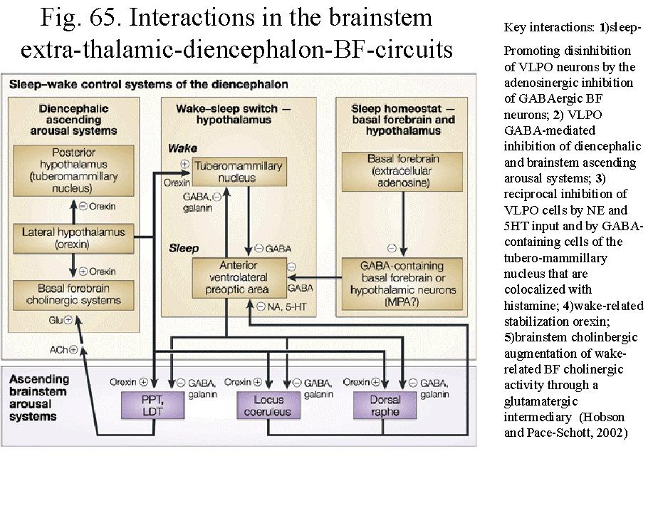
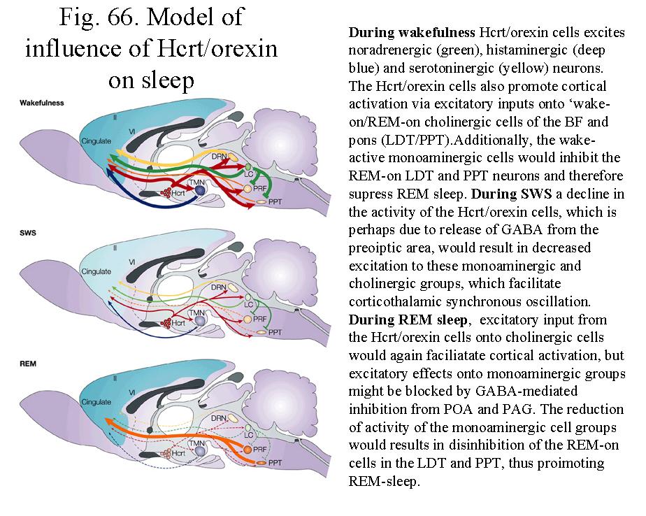
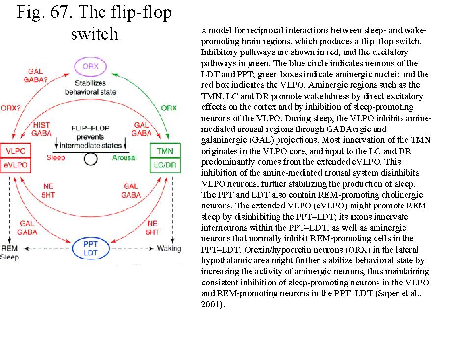
Figs. 65-66. are schematic diagrams to explain the spatial and temporaql interactions in various modulatory systems of the brainstem and sub-thalmic diencephalons/basal forebrain. Figure 67 is a recent model of Saper suggesting the flip-flop switch mechanism of forebrain circuits and their stabilization by the orexin/hypocretin cells of the lateral hypothalamus.
Further Reading
Buzsaki and Traub: Physiological basis of EEG activity. In: Epilepsy: A comprehensive
textbook. Engel et al (eds), Liponcott, 1997
Detari et al: The role of the basal forebrain neurons in tonic and phasic activation of the
cerebral cortex. Progr. Neurobiol, 58, 249, 1999
Dringenberg and Vanderwolf: Involvement of direct and indirect pathways in
electrocorticographic activation, Neurosci and Biobeh. Rev. 22, 243, 1998
Duque, Balatoni, Detari and Zaborszky: EEG correlation of the discharge properties of
identified neurons in the basal forebrain. J. Neurophysiol, 84, 1627, 2000
Hobson and Pace-Schott: The cognitive neuroscience of sleep: Neuronal systems,
consciousness and learning. Nature Reviews Neuroscience, 3, 679, 2002
Jones and Muhlethaler: Cholinergic and GABAergic neurons of the basal forebrain:
Role in cortical activation: Handbook of Behavioral State Control, Lydic Baghdoyan (eds) CRC, 1999
McCormick and Bal: Sleep and arousal. Annu. Rev. Neurosci., 20, 185, 1997
Pace-Schott and Hobson: The neurobiology of sleep: genetics, cellular physiology and
subcortical networks. Nature Review Neuroscience, 3, 591, 2002
Zaborszky and Duque: Local synaptic connections of basal forebrain neurons. Behav.
Brain Res. 115, 143, 2000