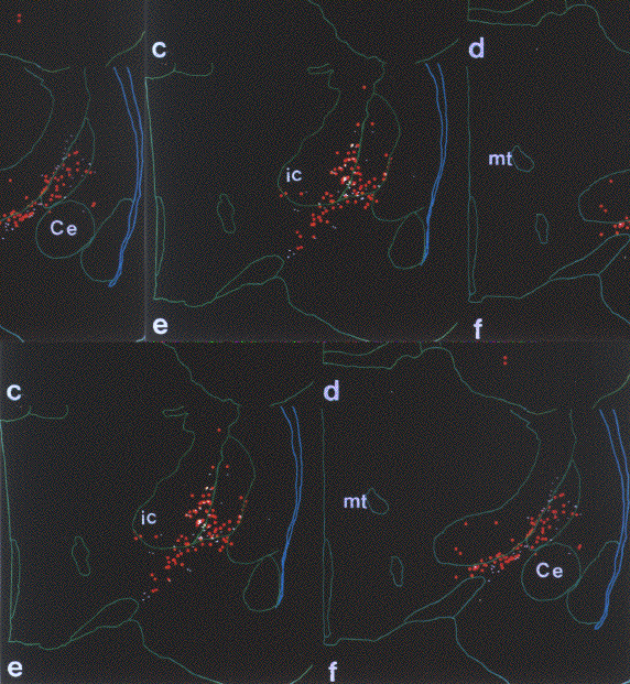
Figure 1.15.1.1 Distribution of single and double labeled cholinergic neurons at six levels in the basal forebrain. Sections were mapped using the Neurolucida image analysis system. For better visualization, the double labeled neurons (ChAT+mGluR) are marked with large red symbols. The "layer" of single labeled cholinergic neurons represented by small white dots are superimposed on the "layer" containing the red neurons and the outlines of the sections.


