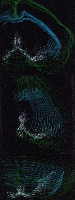
Figure 1.7.-3 Computer graphic reconstruction of cholinergic projection neurons using 12 coronal sections stained for choline acetyltransferase. (NEUROLUCIDA software package). White marks represent cholinergic cell bodies in this and subsequent figures. A: rostral view, B: lateral view, C: dorsal view.


