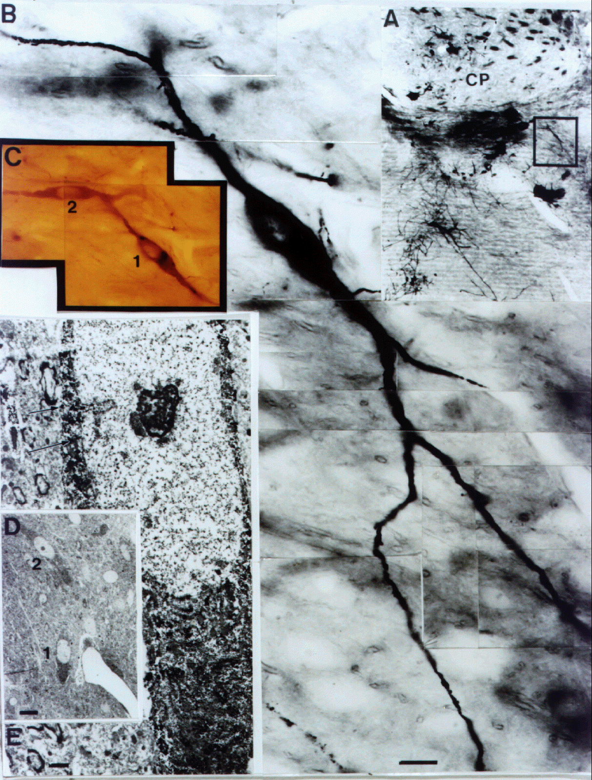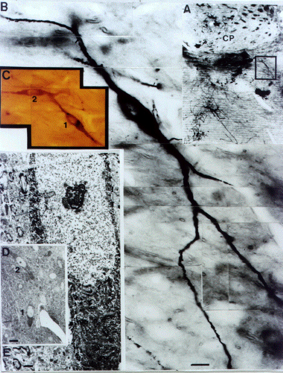
Figure 1.7-4 A: Golgi impregnated cholinergic neuron (method of Gabbott and Somogyi, l984) located medial to the substriatal gray. Neuron in boxed area is shown under higher magnification in B and C. D: Electron micrograph showing gold deposits (arrows) in the peri- karyon, in addition to heavy immunostaining for ChAT. E: Low mag- nification view of the identified neuron (# 1) in the electron microscope. Abbrev. CP = caudate putamen. Scale: B=10”m; E=2”m.