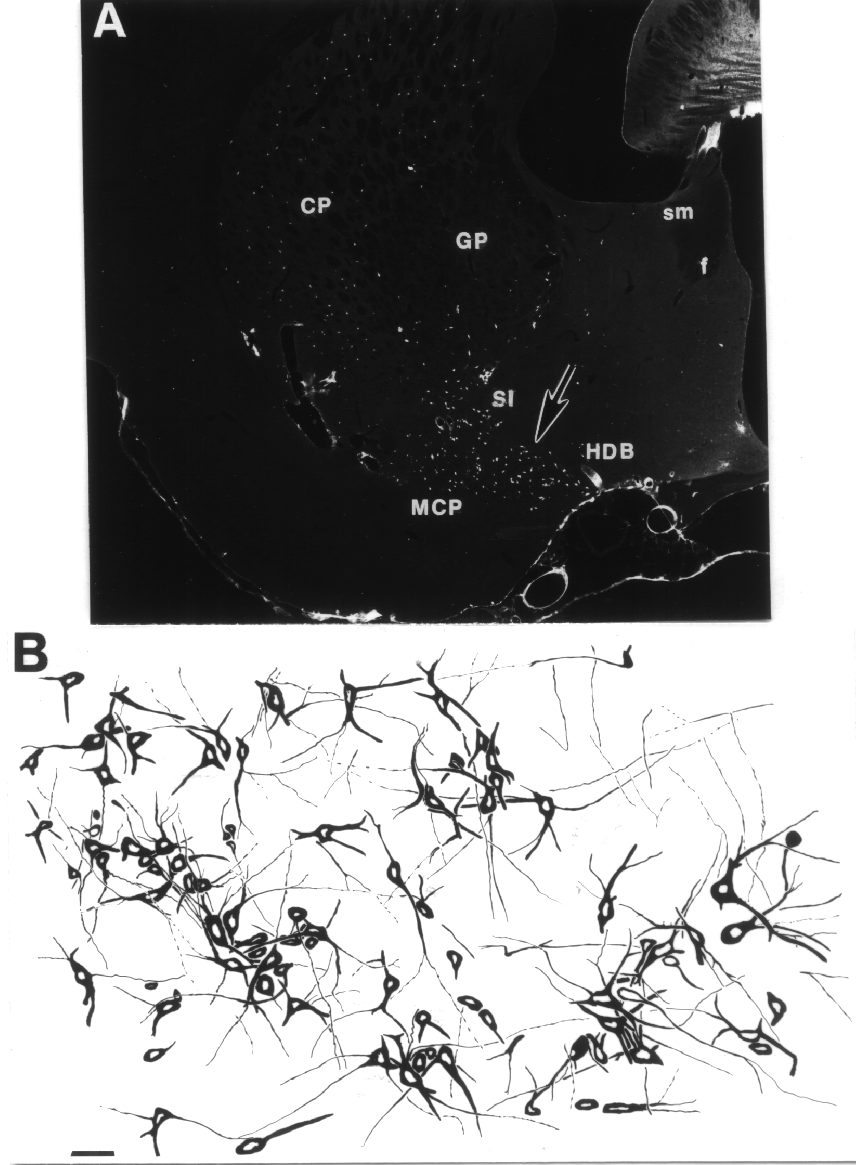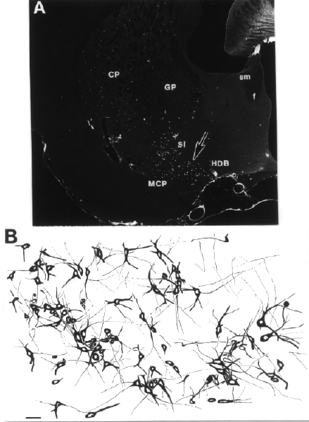
Figure 1.7.5 Distribution of cholinergic neurons in a frontal section of the rat brain approximately 1 mm behind the bregma, depicted by a low magnification darkfield photomicrograph of this area (A). Individual cholinergic cell bodies appear as white dots at this magnification. The section was immunostained with an antibody against choline acetyl transferase (ChAT). GP, globus pallidus; HDB, horizontal limb of the diagonal band; f, fornix; stria medullaris. B Camera lucida drawing from the dorsal part of the HDB from an adjacent section. Approximate location of this area corresponds to the region indicated by an arrow in (A). Note that cholinergic cell bodies often form clusters. Scale: A: 1 mm; C: 50 ”m.