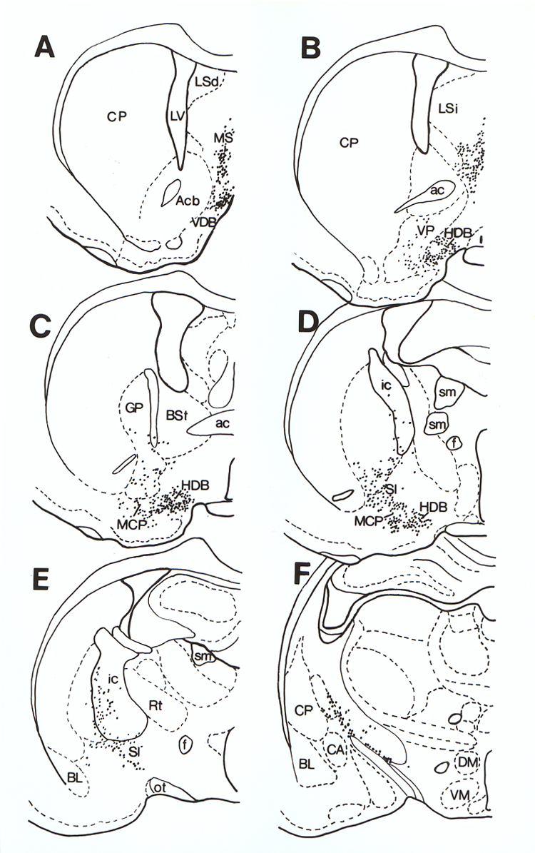
Fig. 1.
A-F: Series of drawings made from
coronal sections through a rat brain (rostral to caudal) that have been immunostained
for ChAT (choline acetyltransferase), illustrating the distribution of
cholinergic neurons (dots). Striatal cholinergic neurons (including those in
the ventral striatum) have been omitted for simplicity. Abbrev. ac =
anterior commissure; Acb = accumbens nucleus; BL = basolateral amygdaloid
nucleus; BSt = bed nucleus of the stria
terminalis; CA = central amygdaloid nucleus; CP = caudate putamen; DM =
dorsomedial hypothalamic nucleus; f = fornix; GP = globus pallidus; HDB = horizontal limb of the diagonal band;
ic = internal capsule; LSd = lateral septal nucleus, dorsal; LSi = lateral
septal nucleus, intermediate; LV =
lateral ven-tricle; MCP = magnocellular preoptic nucleus; MS = medial septal
nucleus; ot = optic tract; Rt = reticular thalamic nucleus; SI = sublenticular
substantia innominata; sm = stria medullaris; VDB = vertical limb diagonal band
nucleus; VM = ventromedial hypothalalamic nucleus; VP = ventral pallidum.