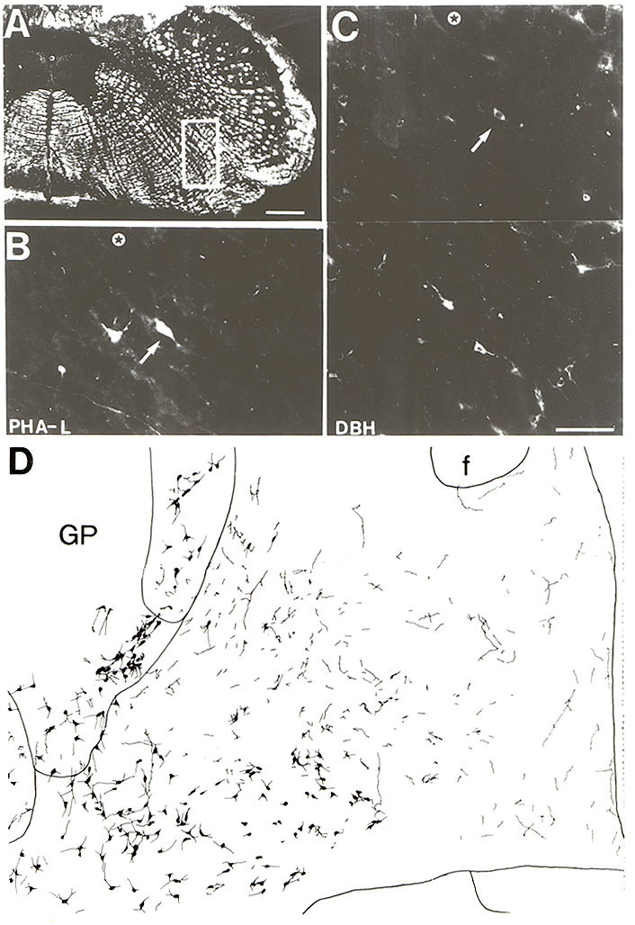
Fig. 11.
A: PHA-L injection site in the
ventral medullary reticular formation. B: Enlarged view of the upper
part of the box of A showing several PHA-L labeled neurons. Arrow points to a
neuron which is double-labeled for dopamine-▀-hydroxylase (DBH). C: The
same section as in B stained for DBH. A number of noradrenergic cells of the A1
group are visible in the lower half of the picture. D: Combined
PHA-L/ChAT staining from the same case showing the distribution of PHA-L
labeled fibers in the forebrain in relation to cholinergic neurons. Bar Scale;
A = 500Ąm; C = 100Ąm. Star indicates the same vessel in B and C. Abbrev.
f = fornix; GP = globus pallidus.