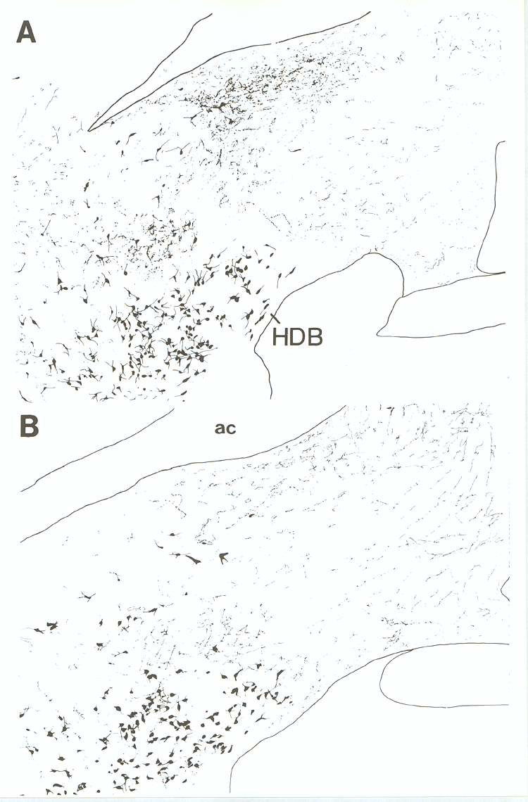
Fig. 13.
A: Camera lucida drawing from a
frontal section at the level of the anterior commissure stained for
dopamine-fl-hydroxylase (DBH) fibers/terminals and cholinergic neurons using the
nickel enhanced DAB/DAB technique. Only the most proximal portions of dendrites
of cholinergic cells are drawn. B: Distribution of PHA-L labeled fibers
in relation to cholinergic neurons at the same level as A after PHA-L injection
in the locus coeruleus. Note that PHA-L labeled fibers/terminals from locus
coeruleus may represent a portion of those found in the DBH/ChAT material. Abbrev. ac = anterior commissure; HDB = horizontal
limb of the diagonal band.