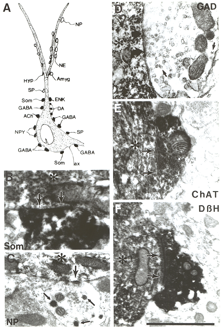
Fig. 14.
A: Topography of synaptic inputs to a
typical cholinergic neuron in the SI. Compiled from our data as well as that of Bolam et al. (l986); Chang et al.
(l987) and Martinez-Murillo et al., (l988a, 1990). Putative inhibitory synapses
are labeled by solid, excitatory contacts by open symbols. B:
Somatostatin-containing bouton in synaptic connection with the dendrite. Double
labeling with nickel enhanced DAB/DAB. C: Bouton containing large
neurosecretory granula (small arrows) establishing synaptic contact with the
distal dendrite. Single immunostaining for ChAT. D: GABAergic bouton
labeled with ferritin (small arrow) establishes symmetrical contact with the
cholinergic cell body. From the material of Zįborszky et al. (1986) by
permission of Willey and Liss. E: ChAT-positive bouton contacts proximal
ChAT-positive dendrite. Single immunostaining enhanced with cobalt. F:
DBH-positive bouton establishes asymmetric synapse with a cholinergic dendrite.
Double immunolabeling with NiDAB/ DAB. Arrowheads show subsynaptic dense
bodies. In all micrographs large arrows point to the postsynaptic side of the
synapse, asterisks label the immunostained (ChAT) profile. Bar scale for all
micrographs: 1 µm.