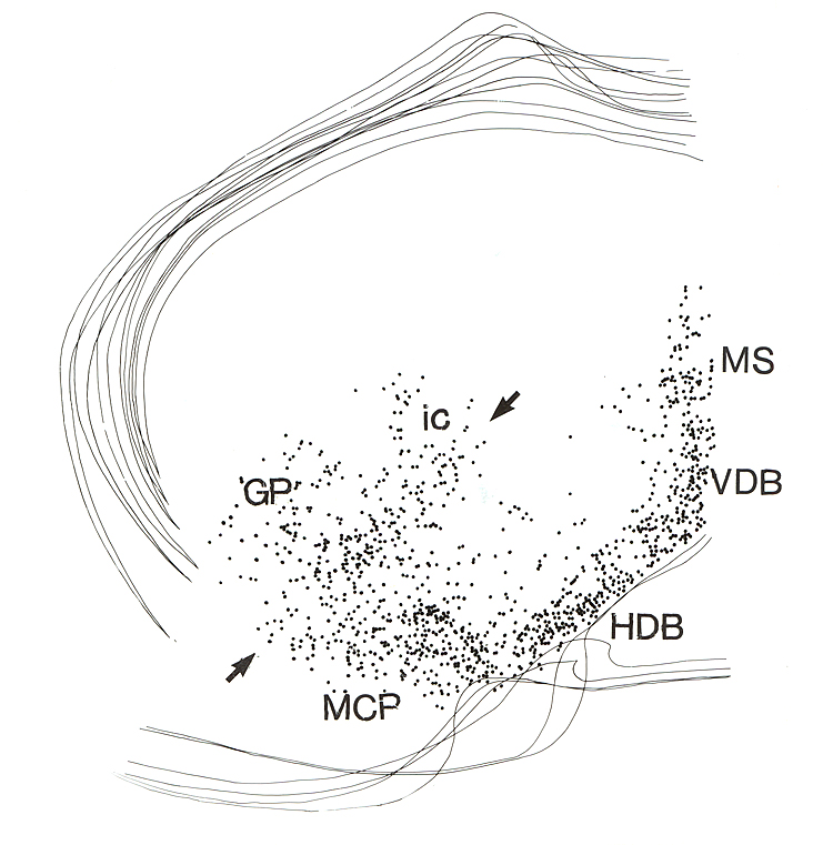
Fig. 2.
Composite map illustrating the distribution of
cholinergic projection neurons (dots). This drawing was composed from 6 camera
lucida drawings which were aligned and superimposed to generate the final
figure. Only the ventral outlines of the brain and the corpus callosum are
indicated. The same technique is used to show the distribution of putative
contact sites of different afferent systems in relation to the cholinergic
neurons in Fig. 15. Cells between the arrows correspond to the area of the SI. Abbrev. ac =
anterior commissure; Acb = accumbens nucleus; BL = basolateral amygdaloid
nucleus; BSt = bed nucleus of the stria
terminalis; CA = central amygdaloid nucleus; CP = caudate putamen; DM =
dorsomedial hypothalamic nucleus; f = fornix; GP = globus pallidus; HDB = horizontal limb of the diagonal band;
ic = internal capsule; LSd = lateral septal nucleus, dorsal; LSi = lateral
septal nucleus, intermediate; LV =
lateral ven-tricle; MCP = magnocellular preoptic nucleus; MS = medial septal
nucleus; ot = optic tract; Rt = reticular thalamic nucleus; SI = sublenticular
substantia innominata; sm = stria medullaris; VDB = vertical limb diagonal band
nucleus; VM = ventromedial hypothalalamic nucleus; VP = ventral pallidum.