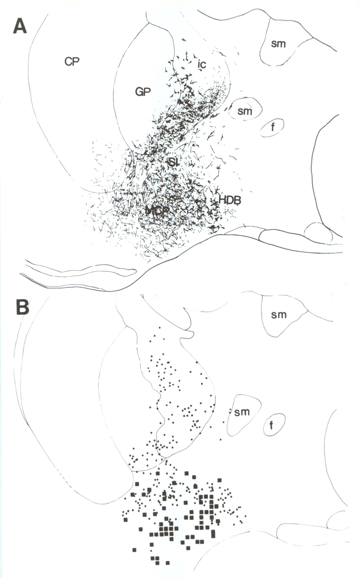
Fig. 4.
A: Camera lucida drawing (10x) from a
double-labeled section illustrating the distribution of PHA-L labeled fibers in
relation to cholinergic neurons following delivery of the tracer to the
orbitofrontal cortex. B: Schematic drawing (from section adjacent to the
one shown in A) illustrating the distribution pattern of PHA-L labeled
terminals in apposition to cholinergic neurons from a section that was analyzed
using high magnification light microscopy. Cholinergic neurons are represented
by dots. Zones of putative contacts between PHA-L-positive terminals and
cholinergic profiles are depicted as squares. Sections were screened using an
ocular reticle (80x80 ”m) at 63x, and contact sites were marked on a camera
lucida drawing of the corresponding section using a proportional grid. Abbrev. ac =
anterior commissure; Acb = accumbens nucleus; BL = basolateral amygdaloid
nucleus; BSt = bed nucleus of the stria
terminalis; CA = central amygdaloid nucleus; CP = caudate putamen; DM =
dorsomedial hypothalamic nucleus; f = fornix; GP = globus pallidus; HDB = horizontal limb of the diagonal band;
ic = internal capsule; LSd = lateral septal nucleus, dorsal; LSi = lateral
septal nucleus, intermediate; LV =
lateral ven-tricle; MCP = magnocellular preoptic nucleus; MS = medial septal
nucleus; ot = optic tract; Rt = reticular thalamic nucleus; SI = sublenticular
substantia innominata; sm = stria medullaris; VDB = vertical limb diagonal band
nucleus; VM = ventromedial hypothalalamic nucleus; VP = ventral pallidum.