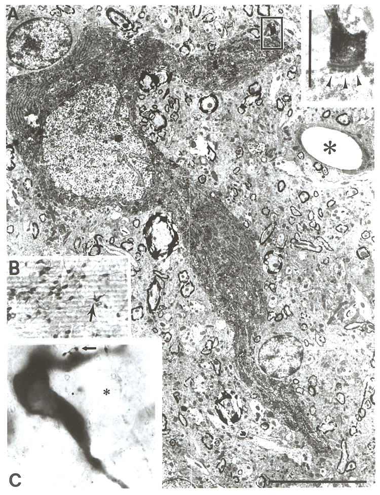
Fig. 7.
A: Low power electron micrograph of a
reconstructed cholinergic neuron from the substantia innominata (arrow in
B). Boxed area in A is enlarged at
inset in upper right corner, and show the synaptic contact of the PHA-L
varicosity, indicated by an arrow in C. Arrowheads in the inset denote sub-synaptic dense bodies. PHA-L was
injected into the lateral hypothalamus. Asterisks in A and C mark the same vessel
for orientation. Bar scale: A = 10 µm, inset = 1 µm. Reproduced from Z·borszky
and Cullinan (1989) by permission of Elsevier Science Publishers.