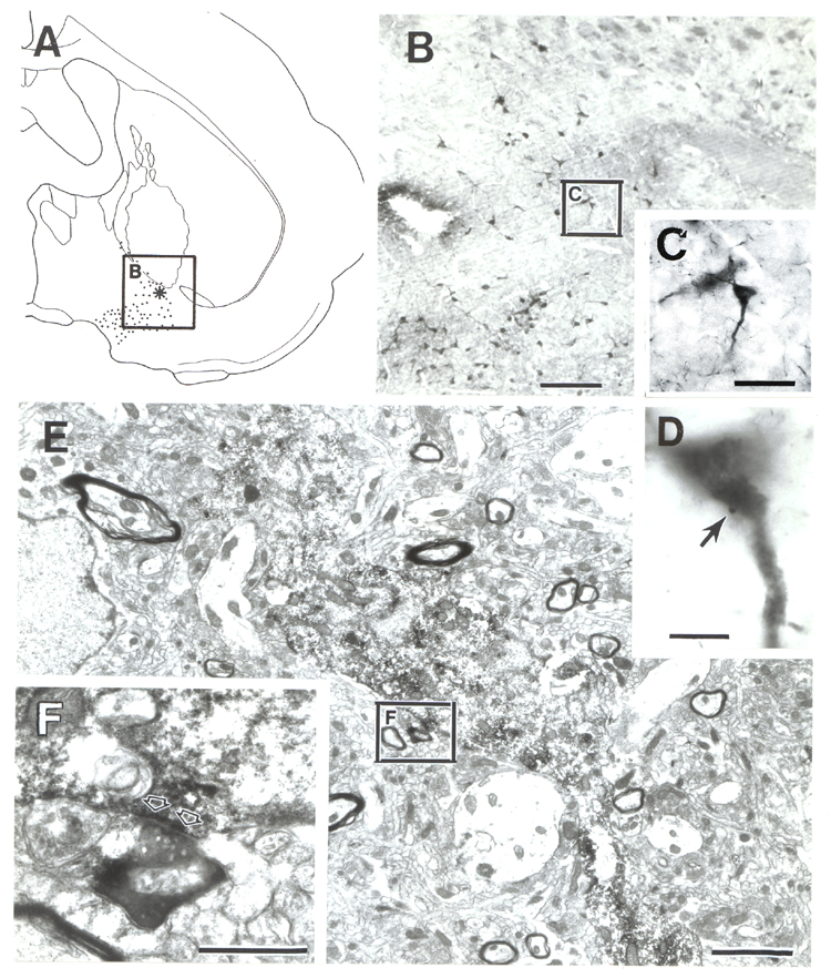
Fig. 10. Micrographs of
afferent input on a proximal dendrite/cell body of a cholinergic cell in the SI
(case 93047). A: Location of the cell indicated by the star in boxed area.
B: Low power micrograph of boxed area in A. ChAT-immunoreactive cell in the center
(in box) is enlarged in C and D. D: Arrow points to the bouton contacting the
dendrite. E: Electron micrograph of the cholinergic neuron. Dendritic shaft,
indicated by electron-dense precipitate, extends to the lower right. Bouton is
present in boxed area F. F: Detail of the strongly electron-dense PHA-L-labeled
terminal. Open arrows indicate the synaptic cleft. Heavy immunoprecipitate at
the synaptic membrane obscures possible postsynaptic thickening. Scale bars =
200 µm in B, 50 µm in C, 10 µm in D, 2 µm in E, 0.5 µm in F.