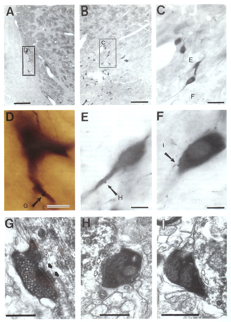
Fig. 11. Examples of nigral innervation of
cholinergic neurons in the basal forebrain. A: Cholinergic neuron in the
lateral part of the BST (boxed area) receiving afferent input from lateral
VTA/medial SNC (case 93047). B: ChAT-immunoreactive cells in GP-SI area
receiving input from the central part of SN (case 93049) with several
cholinergic cells in box C. C: Enlarged view of two neurons in B. The dorsal
neuron is shown at higher magnification in E, and the ventral neuron is
enlarged in F. D: Enlarged view of the neuron from the boxed area in A. Note
the color difference between the cholinergic neuron (brown) and the NiDAB-labeled bouton (black).
E,F: Enlarged view of the boxed areas in C. G: This
bouton establishes an asymmetrical synaptic contact (postsynaptic thickening
indicated with filled arrows). H, I: Electron micrographs of labeled boutons,
shown in E and F, respectively, establishing synapses (open arrows) with
ChAT-positive proximal dendritic shaft (H) and cell body (I). Scale bars = 200 µm in A-C,
10 µm in D-F, 0.5 µm in G-I.