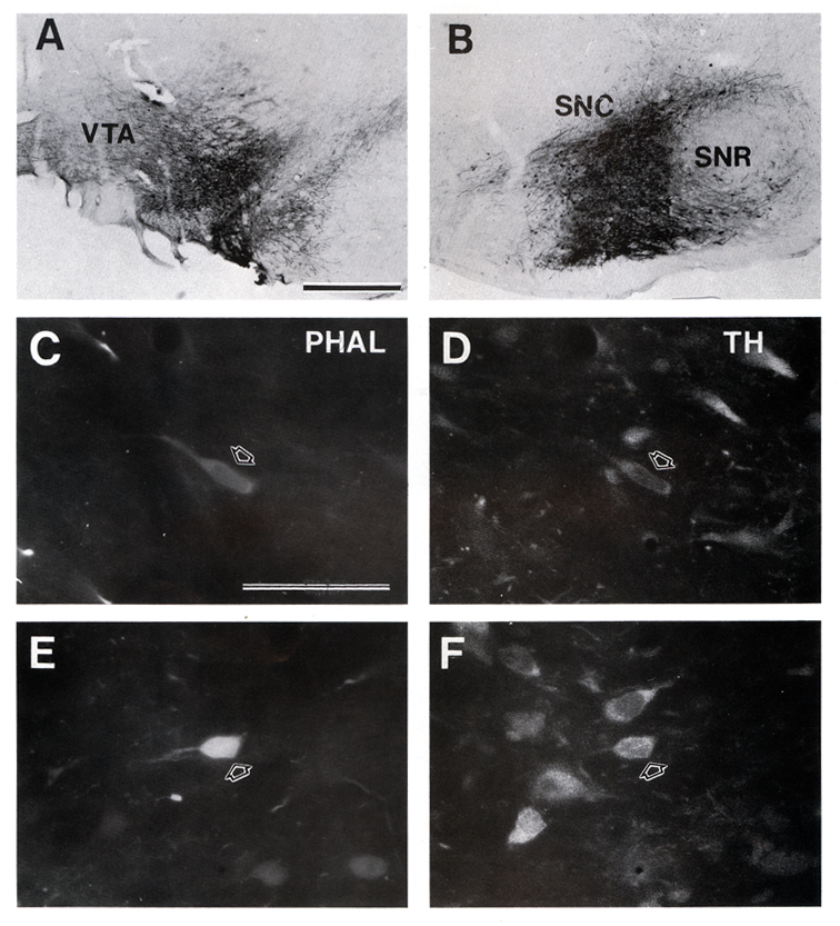
Fig. 2. Photomicrographs of PHA-L injection sites. A:
Tracer-filled cells covering the lateral portion of the VTA and ventromedial corner of the SNC (case 93047).
B: PHA-L-labeled neurons in the central part of the SN (case 93049). C-F:
Double immunofluorescence labeling of PHA-L (Texas Red, C and E) and TH (fluorescein, D and F) within the SNC
(C,D) and VTA (E,F). A large proportion of the PHA-L labeled cell bodies contained TH within the dopamine
cell-rich areas. Scale bars = 50 µm in A (also applies for B), 50 µm in C (also applies for D-F).