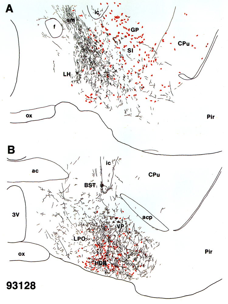
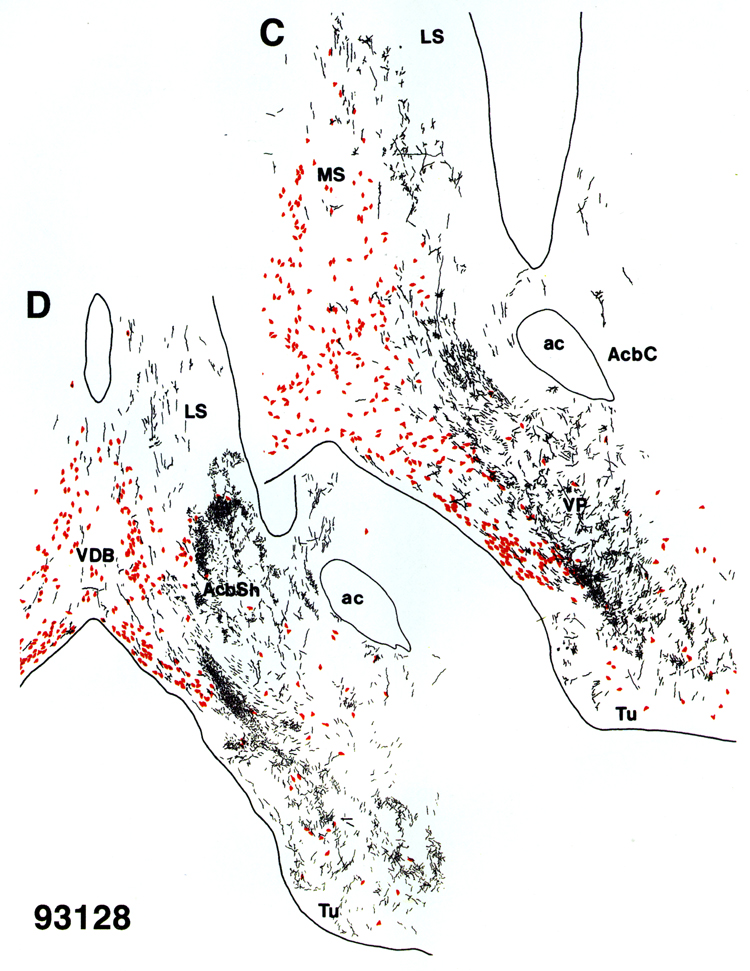
Fig. 4. Camera-lucida-assisted line drawings of
PHA-L-labeled projections in the basalforebrain ascending from the medial portion of the VTA (case 93128).
PHA-L-labeled fibers and boutons (in black) are shown in relation to
ChAT-immunoreactive cell bodies (drawn in red) at 4 different
posterior-anterior levels (A-D, respectively). Figures were drawn from sections
processed for PHA-L-ChAT double-labeling immunocytochemistry.