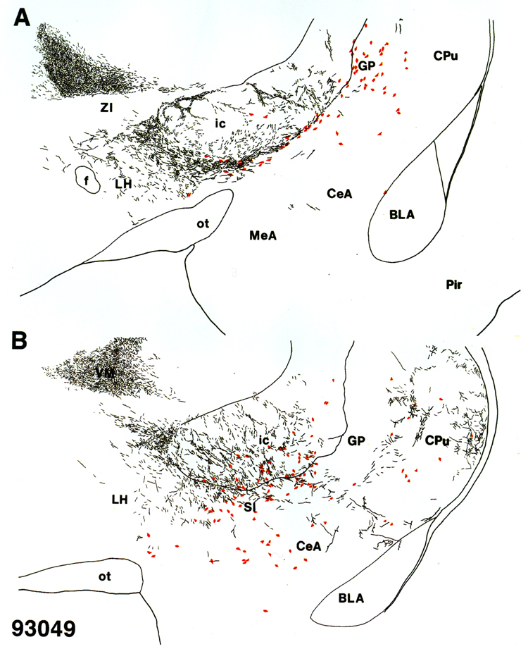
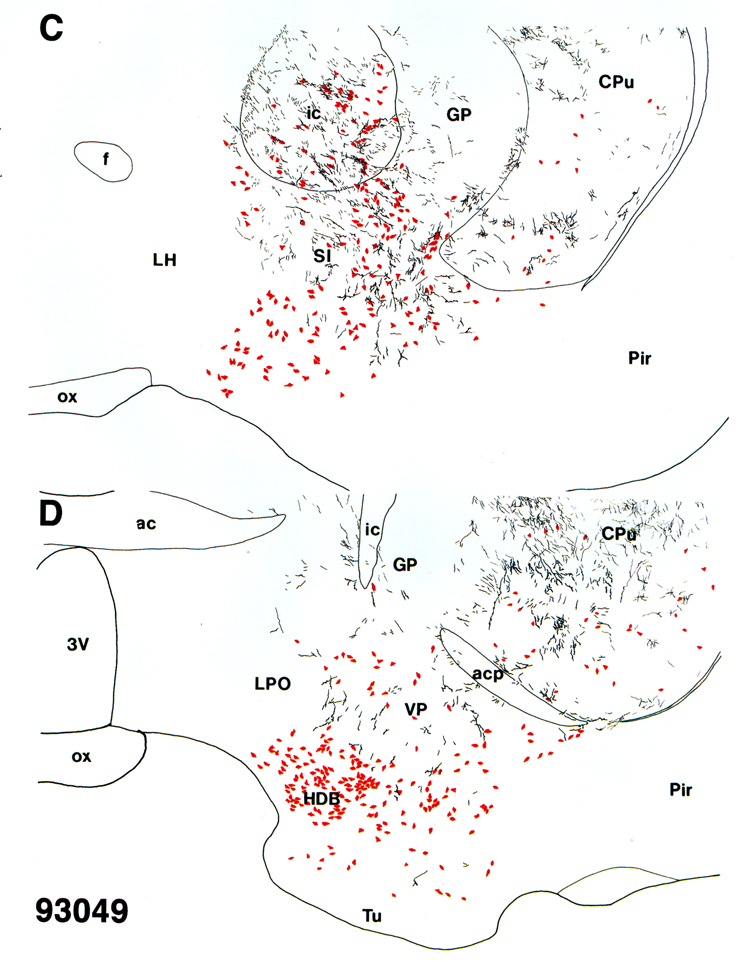
Fig. 6.
A-D:
Pattern of PHA-L-labeled (black) and ChAT-positive (red) structures throughout the basal forebrain of case 93049 with PHA-L deposited in central SN.