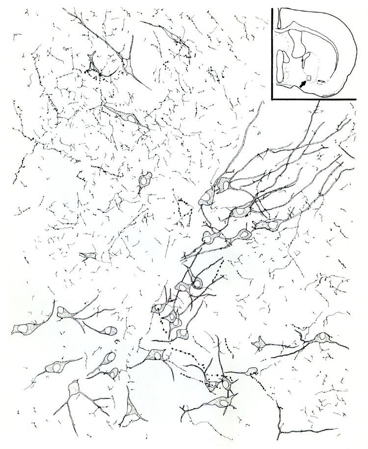
Fig. 2.
Camera lucida drawing illustrating the relationship of cholinergic neurons and
DBH fibers/terminals at the border between substantia innominata and globus
pallidus. The box in the inset shows the approximate location of the drawing.
Note that DBH-positive fibers show different morphology according to the size
and shape of their varicosities and intervaricose segments.