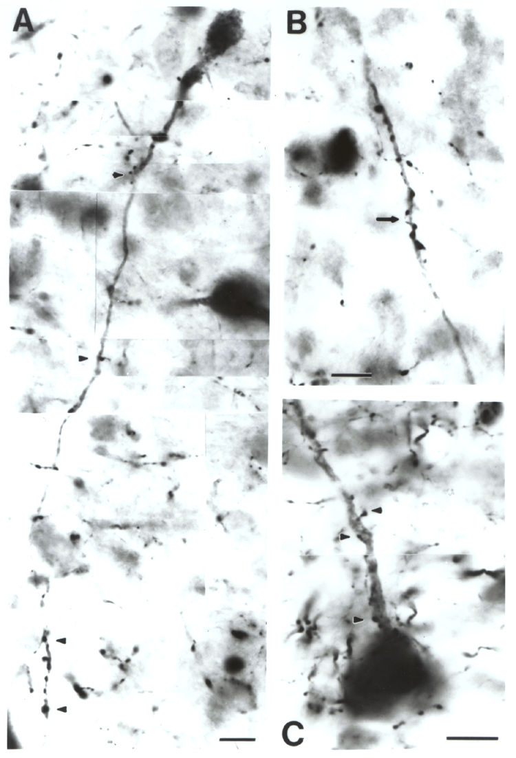
Fig. 3.
High magnification photomicrographs illustrate relationships between
corticopetal cholinergic neurons and DBH (A,B) and TH-positive (C) fibers in
the substantia innominata. A: a cholinergic neuron with cell body in the upper
right corner of this micrograph is approximated by several DBH-positive
varicosities, some of them marked with arrowheads. In the lower left part of the figure the cholinergic dendrite (marked between two arrowheads) is enwrapped by a DBH-positive axon bearing several varicosities. B: a distal cholinergic dendrite is surrounded by at least two axons bearing many varicosities. C: a cholinergic neuron is approached by several TH-positive varicosities. Some of the larger varicosities are indicated by arrowheads. Arrows in B and C point to fine caliber axons. Scale: 10 µm.