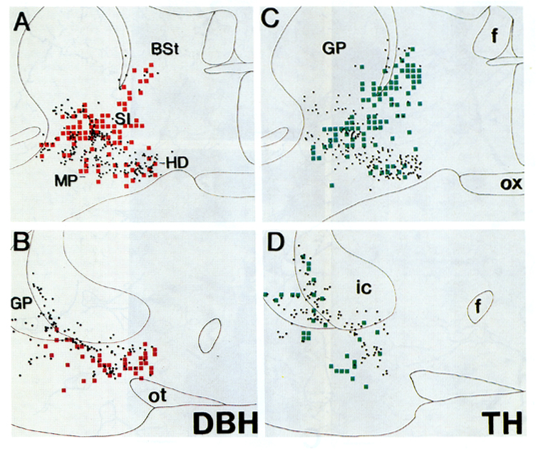
Fig. 4.
Schematic drawing illustrating the pattern of DBH and TH terminals abutting cholinergic
neurons. Alternate sections were processed for DBH/ChAT (A,B) or TH/ChAT (C,D)
double-immunolabeling. Cholinergic cell bodies are represented by black dots.
Zones of putative contacts between cholinergic profiles and catecholaminergic
terminals are depicted as red (DBH) or green (TH) squares. Sections were
screened using an ocular reticle (80 × 80 µm) at 63x and contact sites were
marked on a camera lucida drawing of the corresponding section using a
proportional grid. Putative contact was determined when a clearly identified DBH or TH-labeled terminal (including associated axon) directly abutted a labeled cholinergic profile in the same focal plane. Positive zones generally had 1-3 such arrangements, although occasionally greater number of putative contacts were identified. BSt, bed nucleus of the stria terminalis; f, fornix; GP, globus pallidus; HD: horizontal limb of the diagonal band; MP, magnocellular preoptic nucleus; SI, substantia innominata; ot, optic tract; ox; optic chiasm.