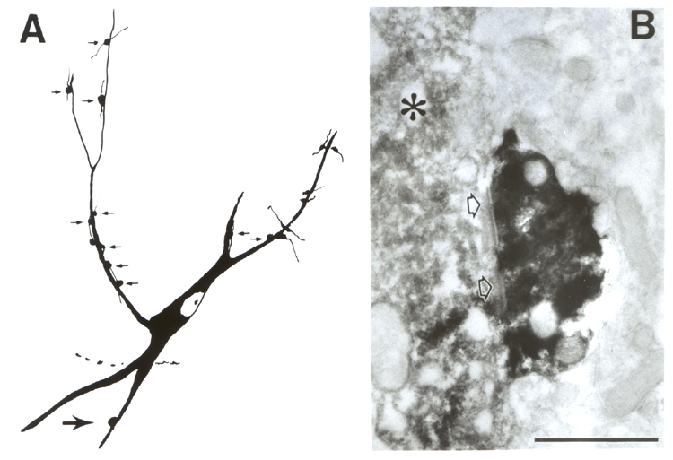
Fig. 5.
A
DBH-positive terminal contacts a cholinergic dendrite. A: Diagramatic
reconstruction of a cholinergic neuron from the substantia innominata,
receiving several varicosities (small arrows) all of which confirmed as synapses by electron microscopy. Large arrow in lower left points to the DBH-bouton shown in B. B: The dendrite of the
cholinergic neuron identified by the flocculent DAB immunoprecipitate is
contacted by a DBH terminal containing the heavy NiDAB deposit. Double labeling
according to Hsu and Soban (l982). Arrowheads denote the postsynaptic
thickening. Scale: 1 µm.