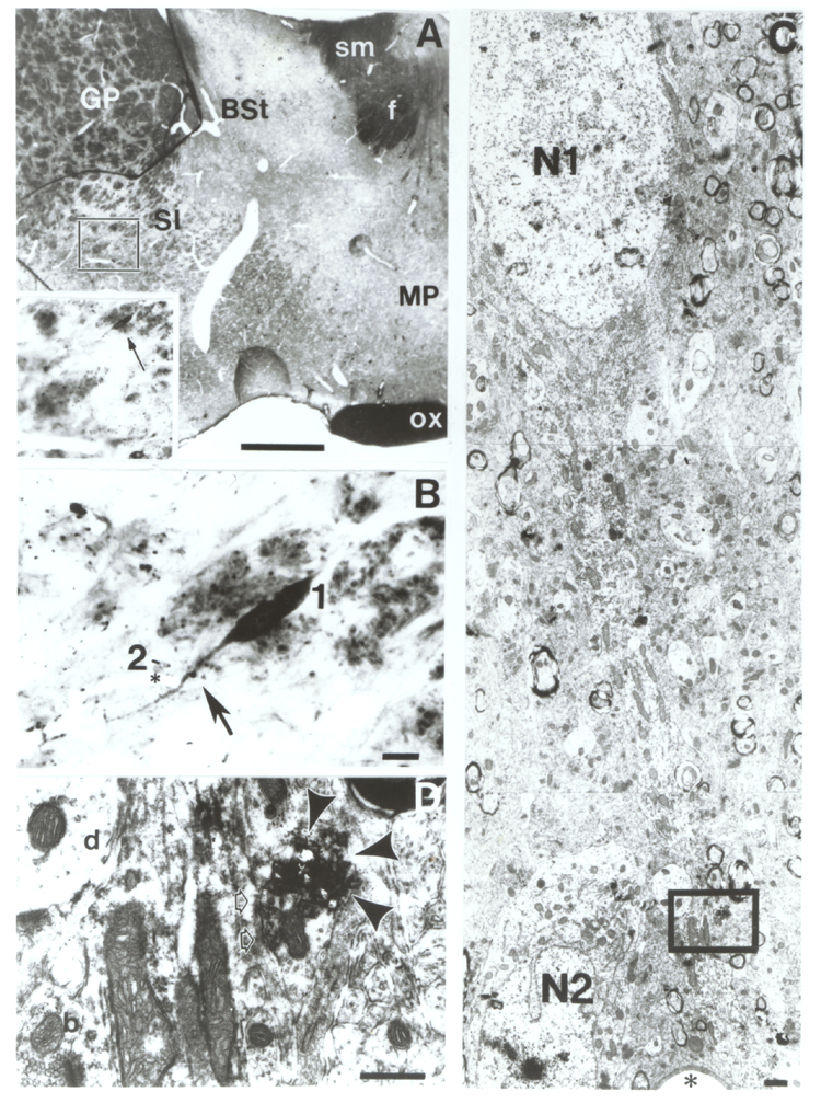
Fig. 6.
Double labeling for TH and ChAT. A: Plastic section of the rostral forebrain
labeled for ChAT and TH using the DAB/benzidine dihydrochloride (BDHC)
technique according to Levey et al., (l986). The boxed area, which is enlarged
in the inset, contains the identified neuron. B: a TH-positive varicosity (arrow) contacts a cholinergic dendrite; the cholinergic neuron (#1) is located among the myelinated fascicles in the
ventral part of the globus pallidus (GP). As fiducial markers #2 denotes
another unlabeled neuron and asterisk indicates a capillary. C: Low power
electron micrograph rotated counterclockwise 45 degrees relative to (B) shows
the same cholinergic neuron (N1= cell body); N2 and capillary (asterisk) serve
for correlating (B) and (C). The framed area in (C) is shown at higher
magnification in (D). D: The dendrite of the cholinergic neuron identified by
the diffuse DAB precipitate is contacted by a TH-positive terminal containing
the heavy BDHC deposit (arrowheads). Open arrows denote the postsynaptic site.
For comparison see an unlabeled dendrite (d) and an unlabeled axon terminal
(b). BSt, bed nucleus of the stria terminalis; f, fornix; ox, optic chiasm; SI,
substantia innominata; sm, stria medullarisScale: A: 0.5 mm, B: 10 µm, C and D:
1 µm.