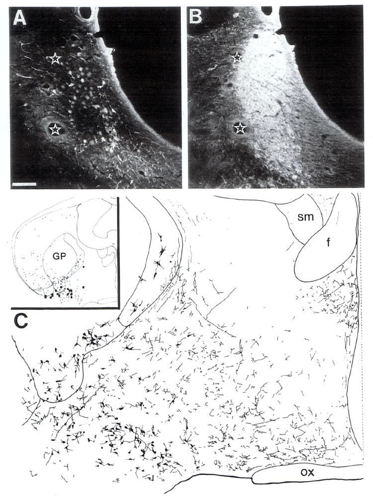
Fig. 7.
Tracing of axons from the locus coeruleus to basal forebrain corticopetal
cholinergic cells. A: PHA-L injection site in the locus coeruleus. B: The same
section immunostained with an antibody against DBH. Double fluorescence
(FITC/RITC) technique. Note that all PHA-L labeled cell bodies are confined to
the heavy DBH-positive area. Stars denote the same vessels. Scale: 100 µm. C:
Camera lucida drawing showing the distribution of PHA-L labeled fibers in
relation to forebrain cholinergic neurons following injection of the tracer
into the locus coeruleus. Inset shows an adjacent section to (C) which was
mapped at high-magnification light microscopy for the presence of putative contacts
between PHA-L labeled varicosities and cholinergic neurons. Cholinergic cell
bodies are represented by dots. Zone of putative contacts between cholinergic
profiles and PHA-L labeled terminals are depicted as black squares
(corresponding 80 × 80 µm in the section). GP, globus pallidus; f, fornix; ox,
optic chiasm; sm, stria medullaris.