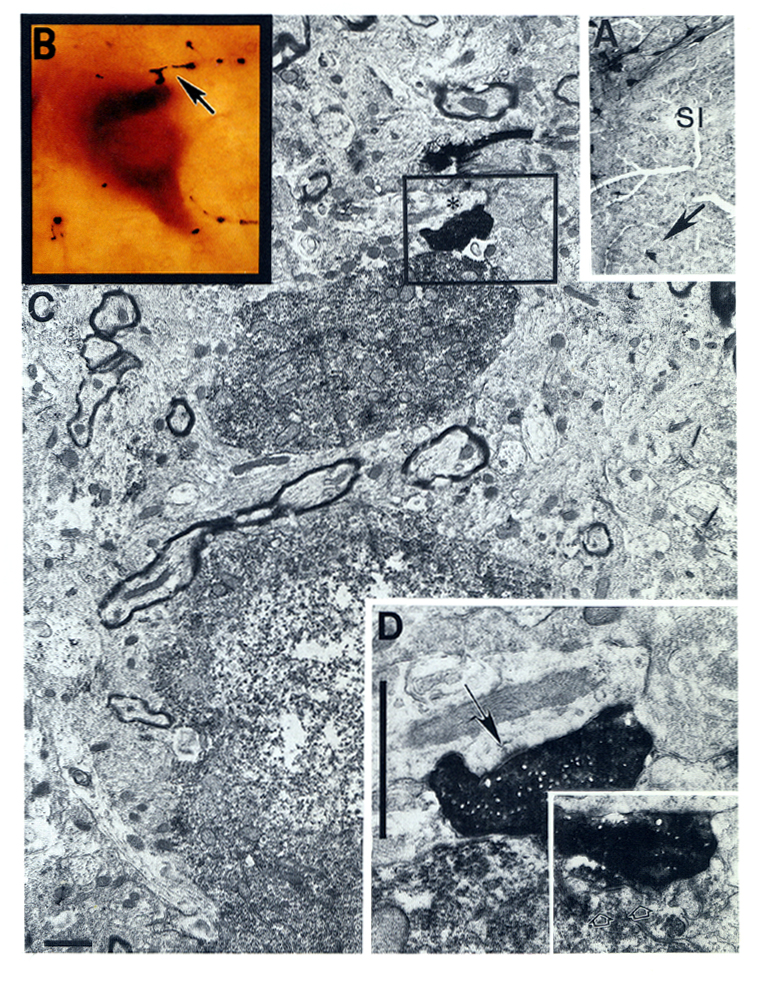
Fig. 9.
Correlated light and EM study to show that identified cholinergic neurons
receive terminals from the locus coeruleus. A: The identified cholinergic
neuron, which is located in the substantia innominata (SI), is approached by a
PHA-L fiber bearing several varicosities (arrow in B). C: Low-power electron
micrograph showing the cell body and part of a dendrite of this neuron. D: An
enlargement of the boxed area from (C) showing that the PHA-L labeled terminal
bouton makes an asymmetric synapse with an unlabeled dendrite (asterisk in C).
Inset from an adjacent thin section demonstrates that the same PHA-L labeled terminal also contact the cholinergic dendrite. Arrows in D and in the inset point to the postsynaptic densities.
Scale: in C and D: 1 µm.