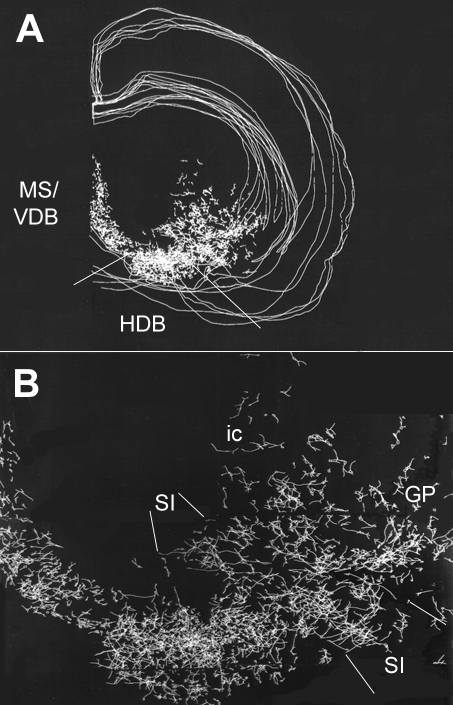
Fig. 1. Composite map illustrating the distribution of cholinergic neurons and their initial (about 150 µ ) dendrites. This map was generated from 7 sections stained for choline acetyltransferase using the Neurolucida software package. For better visualization only the outlines and the corpus callosum are indicated. Anterior view. (A) low magnification, (B) enlargement from (A). Note that orientation of the dendrites show systematic shift along the rostro-caudal continuum of cholinergic cells. Abbr. MS/VDB = medial septum/vertical limb of the diagonal band; HDB = horizontal limb of the diagonal band; GP = globus pallidus; ic = internal capsule; SI = substantia innominata. From the unpublished material of J. Somogyi and Záborszky.