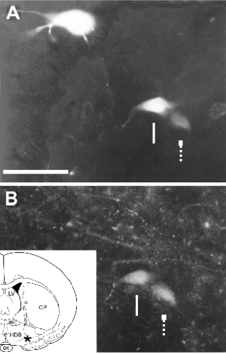
Fig. 7.
Parvalbumin-containing cells projecting to the prefrontal cortex. (A) three Fluoro-Gold labeled cells. (B) Two of the retrogradely labeled cells are positive for parvalbumin (arrows). Lower inset shows the location of the photomicrograph (star). CP = caudate putamen; LV = lateral ventricle, ox = optic chiasm; ac = anterior commissure; HDB = horizontal limb of the diagonal band. Scale bar: 50 µ.