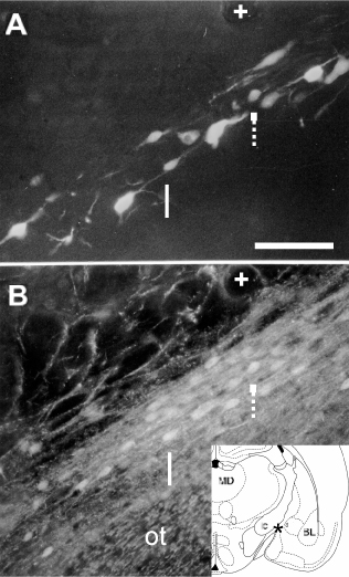
Fig. 9.
Calretinin-containing (CR) neurons projecting to the auditory cortex. (A) Row of retrogradely labeled cells in the narrow zone between the optic tract (ot) and the internal capsule. (B) Two of the CR-containing neurons (arrows) are retrogradely labeled in (A). Lower right inset shows the location of the micrographs. + sign label the same vessel. MD = mediodorsal nucleus of the thalamus; BL = basolateral amygdaloid nucleus; ic = internal capsule. Scale bar: 100 µ.