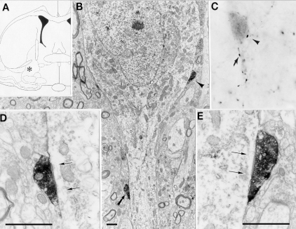
Fig. 11.
Synaptic input from the brainstem to a PV-containing neuron in the ventral pallidum (A: asterisk). (B) Low magnifcation overview of a PV-containing neuron. Arrows point to PHA-L labeled synaptic terminals. (C) high magnification light micrograph of the PV-containing neuron depicted in (B). Arrows point to PAH-L labeled varicosities originating from the injection site in the brainstem (Fig. 10A). (D, E) high magnification electron micrographs of the left and right synaptic boutons from (B). Arrows point to the postsynaptic site. Scale bars: 1 µ.